Why a Vendor-Agnostic IMS Is Key to a Future-Proof AI Strategy
by Patrick Myles, MBA CEO, PathPresenter
As AI adoption accelerates in digital pathology, many institutions are facing a strategic decision that will shape their technology stack for years to come: how tightly should AI be coupled to the core imaging and workflow platform?
At PathPresenter, we’ve taken a very deliberate position. On the AI side, we are intentionally AI-agnostic at the IMS and workflow level. That decision is rooted not in theory, but in hard-earned lessons from the field.
The Hidden Cost of Closed AI Ecosystems
Over the years, we’ve seen multiple institutions make large investments in closed scanning and workflow ecosystems, often driven by early regulatory approvals or the promise of an “end-to-end” solution. In the short term, those systems worked. But over time, they frequently became bottlenecks.
When institutions later wanted to onboard additional scanners, integrate third-party tools, or adopt stronger algorithms from competing vendors, those closed platforms made change slow, expensive, or in some cases impossible. The result was delayed deployments, integration failures, and ultimately costly replacement projects that disrupted operations far more than anyone anticipated.
That model simply doesn’t hold up in today’s AI landscape.
AI Is Changing Too Quickly for Locked-In Platforms
AI is moving at a pace we’ve never seen before. Many algorithms deployed in production today are CNN-based, while the next wave of capability is clearly shifting toward foundation-model approaches. As performance improves and new vendors emerge, the “best” solution will continue to change.
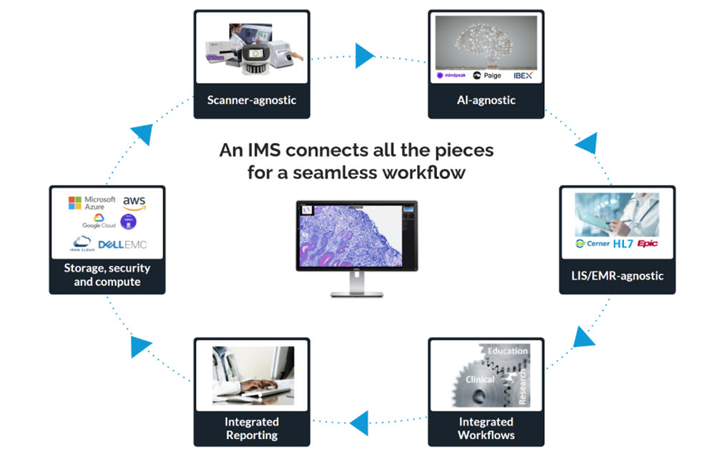
In that environment, locking the core IMS and workflow layer to a specific AI vendor is a strategic risk. Our view is simple: the IMS should remain independent of the AI layer.
A Future-Proof Strategy that Keeps the Platform Stable While AI Evolves
To make that separation practical, we’ve invested heavily in building an AI middleware layer designed to be future-proof. This middleware can ingest, normalize, and orchestrate the clinical and operational data required to deploy AI at scale, including integrations built on HL7, FHIR, and DICOMweb. By abstracting these complexities away from the core platform, institutions can adopt new AI models and workflows over time without needing to rebuild integrations or re-platform each time the ecosystem evolves.
Just as importantly, this approach is informed by real-world experience. We’ve worked with a wide range of institutions and organizations and have seen multiple generations of digital pathology and AI vendors come and go. Some early companies were highly innovative, but struggled to scale or keep pace with market shifts.
Those experiences have consistently reinforced the same conclusion: workflow platforms must remain stable, while the AI layer must remain flexible.
In an AI landscape defined by constant change, future-proofing isn’t about predicting which algorithm will win. It’s about designing systems that can evolve, without disruption, as the technology does.
About the Author
Patrick Myles is CEO of PathPresenter. Previously, he was CEO of Huron Digital Pathology, and Vice President of Business Development for Teledyne DALSA. He served as a board member of the Digital Pathology Association.
Why Seamless IMS-LIS Integration Is Essential for Digital Pathology
Unified digital workflows connecting IMS to LIS unlock efficiency, compliance, and the full potential of AI for modern pathology
Digital pathology has moved decisively from experimentation into everyday practice. For pathology organizations, it now underpins clinical workflows, operational efficiency, and long-term strategy. To enable remote sign-out and AI-driven analysis, organizations have turned to Image Management Systems (IMS) that can organize, track, and contextualize whole slide images across the diagnostic workflow.
However, the data needed to interpret those images extends well beyond what an IMS typically manages. Patient demographics, clinical context, specimen metadata, test orders, results, finalized reports and more all live in a separate but equally critical system: the Laboratory Information System (LIS). For groups accustomed to thinking primarily in terms of image workflows, the LIS can feel like a parallel universe, yet it is the system of record for nearly all non-image pathology data. When IMS and LIS platforms operate in isolation, inefficiencies emerge, context is lost, and the promise of digital pathology remains only partially realized.
As digital pathology programs mature, it is becoming evident that success depends not only on performance, scale, and regulatory readiness, but also on seamless interoperability. In this article, we explore why deep, context-aware integration between an IMS and a modern LIS is essential to unlocking the full value of digital pathology. We’ll examine the practical challenges this integration resolves and the strategic benefits it enables for pathology groups advancing toward a fully digital, AI-enabled future.
Image Management Systems (IMS) in Digital Pathology
A digital pathology image management system (IMS) goes far beyond mere image viewing. At its best, it is an enterprise-grade platform designed to manage the entire lifecycle of digital pathology images across clinical, research, and educational workflows, and serving as the backbone of digital pathology operations, supporting everything from secure storage to AI integration and multi-institution collaboration.
Core Capabilities of a Modern Digital Pathology IMS
A robust IMS typically includes:
Advanced Slide Viewing: High-performance visualization across multiple formats (SVS, NDPI, SCN, DICOM, BigTIFF, and more), synchronized multi-slide viewing, annotations, overlays, and fast rendering.
Centralized Data Management: Secure, scalable image storage, on-premises, cloud, or hybrid, with governance controls and long-term data stewardship.
Security and Compliance: HIPAA-grade protections, encryption, audit trails, role-based access, and regulatory readiness for clinical use.
Workflow Integration: Case management, task tracking, and seamless interoperability with LIS, EHR, PACS, and other enterprise laboratory software systems.
User Roles and Access Control: Granular permissions ensure that only authorized users access sensitive data, while maintaining accountability.
Collaboration Tools: Support for remote consults, tumor boards, education, and peer review through shared environments.
AI and Image Analysis Enablement: Integration with AI models for detection, quantification, and pattern recognition, embedded directly into diagnostic workflows.
Regulatory Readiness: Many IMS platforms are designed to meet FDA, HIPAA, GDPR, IVDR, and other regulatory standards. Some, such as PathPresenter’s clinical viewer, are FDA 510(k) cleared and EU-IVDR certified for primary diagnosis when paired with approved scanners.
In short, an IMS provides a comprehensive infrastructure for digital pathology. It can be highly capable on its own, handling slide storage, visualization, annotations, and even AI outputs. But when it operates independently from the laboratory’s core operational system, its impact is inherently limited. Rather than dissolving legacy barriers, a disconnected IMS can unintentionally recreate them in digital form.
True digital pathology maturity is reached only when images and laboratory data function as a single, coherent workflow. Without that alignment, pathology groups often find themselves managing parallel systems that never quite converge.
The Operational Cost of Keeping IMS and LIS Separate
When image workflows and laboratory workflows are not connected at a foundational level, pathology teams encounter familiar but amplified challenges, including:
Reliance on manual reconciliation of cases and slides
Repeated entry of patient and specimen data
Frequent toggling between systems during case review
Greater exposure to mismatches, omissions, and reporting errors
Disjointed compliance records spanning multiple platforms
Collaboration slowdowns across sites and subspecialties
In isolation, each issue may seem manageable. At scale however, particularly in regulated, high-throughput environments, they compound rapidly, eroding both efficiency and confidence in the digital workflow.
Rethinking the LIS: More Than a Back-End Database
For many digital pathology teams, the LIS is perceived primarily as a static repository: the place where orders originate and reports ultimately land. While that historical role remains essential, it no longer reflects how modern laboratories operate.
From Passive Record-Keeping to Active Workflow Orchestration
Today’s laboratories increasingly require systems that do more than store information. A modern LIS such as LigoLab functions as an operational engine: driving workflow logic, enforcing rules, automating handoffs, and presenting the right context at the right moment.
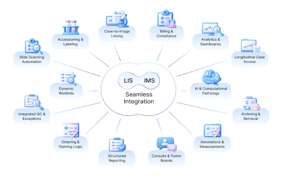
When an IMS is natively integrated into this environment, digital pathology is no longer an external tool that users “visit.” Instead, image review becomes a natural extension of the laboratory workflow, governed by the same logic, permissions, and traceability as the rest of the case lifecycle.
What Integrated LIS–IMS Workflows Enable Day to Day
In a genuinely unified digital pathology environment:
Digital slides are opened directly from the laboratory case, not searched for separately
Patient, specimen, and order context flows automatically into image review
Movement between data, images, and reports feels continuous rather than segmented
Auditability covers both diagnostic decisions and image interactions end to end
AI outputs appear within established workflows rather than as side-channel tools
This level of integration changes how work is performed: not by adding features, but by removing friction.
Core Challenges Solved by Deep Integration
1. Smoother, Faster Diagnostic Workflows
When image access is embedded within the laboratory system, pathologists spend less time navigating software and more time interpreting cases. Reduced friction translates directly into faster turnaround and lower cognitive load.
2. Scalable Support for Remote Practice
Integrated platforms make location largely irrelevant. Secure remote sign-out, real-time collaboration, and distributed subspecialty review become standard operations rather than exceptions.
3. Better Decisions Through Complete Context
Images rarely tell the full story on their own. Tight coupling between LIS and IMS ensures that diagnostic interpretation always occurs alongside the complete clinical and specimen context, reducing the risk of incomplete assessment.
4. Stronger Governance and Compliance
When images and laboratory data share a unified audit framework, compliance with regulatory and accreditation requirements becomes simpler and more defensible—without added administrative burden.
5. A Practical Path to Enterprise AI
AI delivers value only when it operates inside real workflows. Integrated LIS–IMS environments provide the governance, data access, and workflow hooks required to deploy AI responsibly and at scale.
Integration in the Real World: From Concept to Practice
A practical example of this approach can be seen in the collaboration between PathPresenter and LigoLab. Rather than treating image management as an external system, PathPresenter’s clinical viewer is embedded directly within LigoLab’s enterprise laboratory platform.
The result is a unified diagnostic experience where pathologists interact with slides in the same environment where cases are managed, orders are tracked, and reports are finalized. This tight coupling reduces navigation overhead, accelerates review cycles, and establishes a solid foundation for future AI-driven enhancements.
Building Toward a Unified Digital Pathology Future
At its core, LIS–IMS integration is not about software convenience. It is about redefining how pathology work flows from accessioning through diagnosis and reporting.
When laboratory data and images operate as one system:
Operations gain visibility and automation
Financial teams benefit from cleaner alignment with billing and revenue workflows
Clinical staff gain modern tools that reduce manual effort
Patients benefit from faster, more consistent diagnostic outcomes
As digital pathology, AI, and remote diagnostics continue to evolve, this alignment moves from “nice to have” to mission critical. Pathology groups that invest early in deep, contextual integration position themselves not just to digitize slides, but to fundamentally modernize how pathology is practiced.
More Information
Looking for a new (or improved) LIS-IMS integration? Connect with our team to assess your workflow needs and explore the best solution.
Digital Pathology Basics: Image Viewers vs. Image Management Systems
In brief
An image viewer focuses on the basics of viewing and interacting with digital slides.
A digital pathology IMS manages the entire lifecycle of digital pathology images within a clinical, research, or educational workflow.
As digital pathology becomes more integrated into clinical, academic, and research settings, it’s important to distinguish between the tools used in this domain. Two common types of software encountered are pathology slide viewers and digital pathology image management systems (IMS). While they may appear similar on the surface (both enable users to access and interact with digital slides) they are fundamentally different in terms of scope, features, and intended use.
Pathology Slide Viewer: A Focused Viewing Tool
A pathology slide viewer is a software application designed primarily for visualizing digitized pathology slides, also known as whole slide images (WSIs). These tools typically allow users to open and navigate high-resolution scans of tissue specimens. Common features include zooming, panning, rotation, and the ability to add simple annotations or measurements.
These viewers can be used by pathologists for reviewing cases, by students for learning, or by researchers for image analysis. They may be standalone desktop applications or web-based platforms. Some viewers are vendor-specific, tied to a particular scanner or image format, while others are open source or multi-format compatible. However, pathology slide viewers are limited in scope. Their main purpose is to enable manual, visual inspection of slides. They typically do not provide broader workflow functionality, user management, or system integrations. For small-scale or individual use, slide viewers are handy tools. But their limitations will quickly become apparent.
Digital Pathology Image Management System (IMS): A Comprehensive Platform
In contrast, a digital pathology image management system (IMS) such as PathPresenter is a comprehensive, enterprise-grade software solution that manages the entire lifecycle of digital pathology data. It includes not only the viewing of whole slide images, but also the organization, tracking, analysis, and sharing of those images in clinical and research workflows. An enterprise deployment of an integrated digital pathology workflow will almost certainly require a fully featured IMS.
A robust IMS typically includes the following core capabilities:
Slide Viewing: IMS platforms provide high-quality visualization with features like synchronized multi-slide viewing, annotation, overlays, and fast rendering across many file formats (svs, ndpi, scn, dcm, bigtiff, etc.).
Data Management: IMS centralizes and secures pathology data with on-premise, cloud, or hybrid storage options.
Security: IMS must meet strict standards (e.g., HIPAA), ensuring PHI confidentiality, integrity, and availability while protecting against threats.
Workflow Integration: Platforms support case management, task tracking, and diagnostic, research, or education workflows, often with integrated security like SSO.
User Roles & Access Control: Permission-based access and audit trails ensure only authorized users can view sensitive data while maintaining accountability.
Interoperability: IMS integrates with LIS, PACS, EHR, and other systems. PathPresenter is vendor-agnostic, with integrations to leading systems such as EPIC Beaker and LigoLab.
Collaboration Tools: Multi-user environments enable real-time sharing, remote consults (e.g., PathPresenter’s ConsultConnect), and team-based diagnosis or education.
AI and Image Analysis: Advanced IMS platforms integrate AI for detection, quantification, and pattern recognition. PathPresenter partners with leading vendors including Paige, Aisencia, Aiforia, MindPeak, Pictor Labs, Primaa, DeepLIIF, DiaDeep, Artera and IBEX to embed models within its workflow. PathPresenter powers the College of American Pathologists’ (CAP) AI Studio, which lets member test drive AI models from multiple vendors in one site with a common UI.
Regulatory Compliance: IMS solutions are generally designed to meet HIPAA, FDA, GDPR, and or IVDR standards depending on the application and jurisdiction. PathPresenter’s clinical viewer is FDA 510(k) cleared and EU-IVDR certified for primary diagnosis with approved scanners.
Key Differences and Use Cases
While both a slide viewer and an IMS allow for digital slide visualization, their purposes differ greatly. A slide viewer is ideal for simple, low-volume, or individual use cases, such as academic review, research projects without extensive collaboration, or basic diagnostic reference. It is generally easy to deploy and use, and may come free of charge or at a low cost.
A digital pathology IMS, on the other hand, is suitable for high-volume, regulated environments such as hospitals, diagnostic labs, and collaborative research institutions. These systems are designed to scale with demand, handle multi-user scenarios, and provide robust governance and compliance. They also support complex workflows involving multiple roles, locations, and datasets.
Choosing between a viewer and an IMS depends on the specific requirements of the organization or individual, whether it’s ease of access and simplicity, or full integration, scalability, and workflow optimization.
More Information
Not sure if a viewer or IMS is right for you? Connect with our team to assess your workflow needs and explore the best solution.