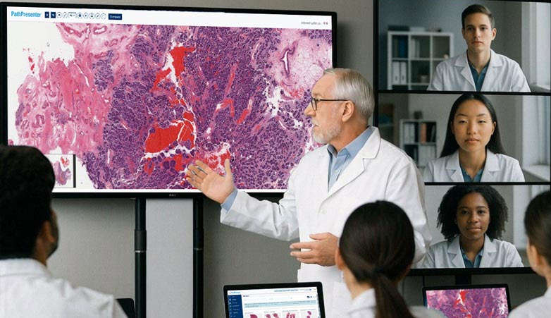
Expanding Access to Hantavirus Education: Sharing Images Through the PathPresenter Public Library
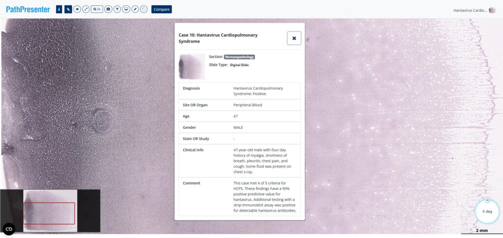
What does hantavirus look like to a pathologist? On a digital whole slide image of a peripheral blood smear, it looks a lot like this.

Hantavirus is a rare but serious infection, and the recent headline-grabbing surge in cases around the world highlights the importance of timely diagnosis in preventing its spread. Recognition of hantavirus may begin with clinical teams seeing symptoms, but pathologists and laboratory professionals play a key role in identifying subtle morphologic features in peripheral blood smears, leading to confirmatory testing and patient management. With this in mind PathPresenter is proud to highlight the work of Dr. Joseph Stenberg, a resident at the University of New Mexico School of Medicine, who is sharing an educational series focused on hantavirus cardiopulmonary syndrome (HCPS) screening in peripheral blood smear cases.

The project comprises 10 hand-selected cases by Dr. Kathryn Foucar, Distinguished Professor at the University of New Mexico, and includes whole slide images (WSIs) representing a broad spectrum of diagnostic presentations. These include classic examples of HCPS, borderline cases, early-evolving infections, and negative mimics that challenge learners to distinguish subtle findings from true disease.

By sharing cases across this diagnostic spectrum, Dr. Stenberg aims to create a valuable educational resource for pathology trainees, practicing pathologists, and other laboratory medicine professionals seeking to improve their recognition of hantavirus-related findings in peripheral blood smears.

Bringing Interactive Learning to Published Medical Education

A key goal of the project is to allow learners to do more than simply view static images. To maximize educational value, Dr. Stenberg is using PathPresenter to provide an interactive slide-viewing experience.

“This project was built around a simple goal: to make high-quality educational cases of hantavirus cardiopulmonary syndrome freely available to anyone, anywhere” said Dr. Stenberg. “By providing unrestricted access to complete digital slides through PathPresenter, we hope to help laboratory professionals become more confident in recognizing this rare but life-threatening syndrome. Earlier recognition can lead to earlier diagnosis, more timely clinical intervention, and ultimately better patient care.”

Rather than relying on selected screenshots or figure panels alone, learners can explore the complete digital slides themselves, zooming and navigating through the specimens just as they would at a microscope. This approach allows users to appreciate subtle findings, evaluate areas of interest independently, and gain experience that more closely resembles real-world diagnostic practice.

To make the cases as accessible as possible, Dr. Stenberg has generously donated de-identified slides to the PathPresenter Public Library. The slides will also be linked from his forthcoming publications (both online and in print), allowing readers to move seamlessly between published educational content and the original digital pathology slides.

A Great Example of Community Contribution

Dr. Stenberg’s project exemplifies one of the core purposes of the PathPresenter public platform: creating a collaborative learning community where educators and learners can contribute to the advancement of pathology education.

Any PathPresenter public user can share de-identified educational material with the community. Contributions from individual pathologists, trainees, academic institutions, and professional organizations collectively help build a richer and more diverse educational ecosystem.

Many users are familiar with PathPresenter’s highly popular High Yield Cases, which are curated and annotated by distinguished pathologists and serve as valuable study resources for residents preparing for board examinations. But the continued growth and success of the platform is driven by contributions from across the pathology community.

Today, the PathPresenter public platform includes more than 85,000 users and over 10,000 slides, representing a broad range of subspecialties, diseases, and educational approaches. Every new contribution helps expand the breadth of learning opportunities available to pathologists around the world.

A Valuable Resource for Hematopathology Education

The HCPS cases contributed by Dr. Stenberg consist of blood smear (hematopathology) images scanned at 40× magnification, providing the resolution needed to examine important cellular details and morphologic features.

Because the collection includes both classic and challenging presentations, it offers learners an opportunity not only to recognize hallmark findings but also to develop diagnostic judgment in cases where the answer may not be immediately apparent. These are often the cases that provide the greatest educational value and most closely reflect real-world practice.

As his cases are published, they will serve as a useful bridge between traditional academic publication and interactive digital pathology education.

Thank You, Dr. Stenberg

PathPresenter salutes Dr. Stenberg and his colleagues for this valuable contribution to publicly available medical knowledge. By sharing these cases with the pathology community, they are helping create resources that will benefit learners and educators far beyond their own institution.

We look forward to sharing links to Dr. Stenberg’s published articles in the near future and will post updates here on the PathPresenter blog as they become available.

In the meantime, if you’d like to explore the slides yourself, visit the PathPresenter Public Library and search for “hantavirus.” If you don’t already have one, you can create a free account which includes 10 GB of storage for your own uploads. 
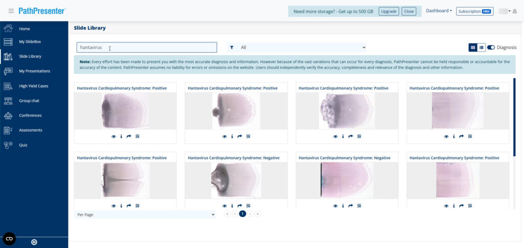
Hantavirus slides in the PathPresenter Public Library

We encourage educators, trainees, and practicing pathologists alike to take advantage of these interactive teaching materials—and to consider contributing their own educational cases to help strengthen the global learning community.

Pathology Education: The Digital Future Is Here

The next generations of pathologists are demanding digital education now

As medicine becomes more precise and data-driven, pathology is undergoing a transformative shift toward digital technology. This transition promises to revolutionize how pathologists work, improving accuracy, efficiency, and collaboration, while ultimately enhancing patient outcomes. Nowhere is this transformation happening faster or more profoundly than in pathology education. And increasingly, it is pathology trainees—residents and fellows—who are driving the demand.

A recent example from the Yale School of Medicine puts this shift into clear focus. When distinguished Yale professor and internationally recognized genitourinary pathologist Dr. Peter A. Humphrey, MD, PhD, retired in 2025, he left behind a spectacular and unique collection of pathology slides that he had used to educate generations of trainees about rare and complex cases. Over 40 years of practice, consultation, and teaching, Dr. Humphrey amassed not just slides, but insight: annotations, diagnostic pearls, and pattern-recognition wisdom refined over decades.

Through a project led by Dr. Roxanne Wadia, these slides are now being digitized (using the PathPresenter platform), along with Dr. Humphrey’s annotations, so that his experience can continue to guide new generations of residents and fellows at Yale and beyond. 

“Dr. Humphrey’s slide collection represents a lifetime of mastery, an extraordinary legacy that deserves to be more than preserved,” said Dr. Raj Singh, professor of pathology and PathPresenter co-founder. “It deserves to be shared.”

This kind of knowledge sharing is admirable, but for today’s trainees, that sharing is not just inspiring, it is becoming expected and even essential. At Yale and many other institutions, digital platforms are no longer viewed as optional enhancements. They are becoming core infrastructure for training pathologists in a world that is already digital.

The Advantages of Digital for Pathology Education

When it comes to delivering pathology education, a digital platform such as PathPresenter offers compelling benefits. But viewed through the lens of trainees, these advantages become even clearer: digital tools directly address the daily challenges residents and fellows face.

Eliminating Physical Constraints

Residents learn pathology by seeing cases—lots of them. Yet traditional training models often limit access. Glass slides are tied to specific microscopes, labs, and physical locations. Rare cases may be archived and difficult to retrieve. Multi-headed scopes allow small-group teaching, but only for those physically present.

Digital platforms eliminate these constraints. With whole-slide imaging, specimens can be digitized at high resolution and shared almost instantly with learners anywhere in the world. Instead of crowding around a microscope, trainees can review cases independently, revisit them after sign-out, and study them before board exams. They can zoom, pan, and explore at their own pace, without worrying that the slide must be returned to circulation.

For busy residents balancing clinical service, conferences, research, and exam preparation, this flexibility is transformative. Learning is no longer limited to “when the slide is available.” It becomes on-demand and continuous.

Pathology depends heavily on visual pattern recognition. Even the most detailed written description of a cellular structure cannot match the power of an image. When trainees and attendings view the exact same digital slide simultaneously, discussions are more precise. Feedback is clearer. Diagnostic reasoning becomes more transparent.

Digital images also preserve rare and valuable cases that trainees might otherwise never encounter. Instead of being stored in a single institutional archive, these collections can be shared globally. For residents preparing for subspecialty fellowships or boards, exposure to rare entities is invaluable.

This is precisely what Yale envisions for Dr. Humphrey’s collection: preserving and sharing his legacy so it can continue to guide and challenge future pathologists. Open access to world-class pathology knowledge is not just a philosophical goal, it directly supports trainee growth and confidence.

Unlocking Interactive Learning

Today’s trainees do not want to passively observe, they want to interact.

Digital platforms enable educators to annotate directly on slide images, highlight key diagnostic features, add measurements, and create guided tours through complex cases. For trainees, these annotations become enduring teaching points that can be revisited long after the live session ends.

Learners can also engage actively, leaving comments, asking questions, and even participating in real-time chats. Instead of fleeting conversations at a microscope, discussions become documented and searchable. Slide metadata, clinical history, and diagnostic commentary can travel with the image, ensuring that context is never lost.

For residents preparing for tumor boards, unknown conferences, or board examinations, this depth of context is critical. It allows them to connect morphology with clinical implications, immunohistochemistry, molecular findings, and outcomes.

Digital platforms also support structured curriculum delivery. Organized modules, case libraries, integrated quizzes, and assessments provide clarity in a training environment that can otherwise feel overwhelming. Immediate feedback helps trainees identify knowledge gaps early. Progress dashboards and certification tracking bring transparency to competency development.

In an era where pathology training varies across institutions, digital platforms help standardize access to high-quality content. For trainees, that consistency means greater confidence, regardless of where they train.

Enabling Global and Remote Learning

Trainees today are part of a global medical community, and digital pathology platforms break down geographic barriers, allowing residents and fellows to learn from experts far beyond their home institutions. A trainee in a resource-limited setting can now access the same high-quality cases and instruction as a learner at a major academic center.

Virtual tumor boards, international conferences, and cross-border case discussions are no longer rare events, they are increasingly routine. For fellows pursuing subspecialty expertise, exposure to diverse case material and global perspectives strengthens both diagnostic skill and professional networks.

For many trainees, remote learning is not a compromise; it is an opportunity, and perhaps even a necessity.

Integrating Artificial Intelligence into Training

The pathologists of tomorrow will practice in environments shaped by artificial intelligence. AI models are already being integrated into clinical workflows and research applications, from image analysis to diagnostic support.

Trainees recognize this shift. They want to understand not only how to interpret slides, but how to evaluate and leverage AI tools responsibly.

Comprehensive digital platforms provide a natural environment for this integration. In a digital workspace, residents can explore AI-assisted analyses, compare algorithmic outputs with their own interpretations, and develop the critical thinking skills necessary to use these tools wisely.

Exposure during training ensures that future pathologists are fluent in both traditional morphology and digital augmentation. Digital literacy is becoming as fundamental as microscopic technique.

Meeting Trainee Expectations

Today’s residents and fellows are digital natives. They expect intuitive interfaces, instant access to resources, and collaborative tools that resemble the platforms they use in other aspects of their lives.

They also understand that their future employers (academic centers, hospitals, laboratories) are moving toward digital pathology workflows. They want their training environment to reflect that reality.

Institutions that embrace digital platforms signal to trainees that they are forward-looking and invested in preparing them for modern practice. Those that do not risk falling behind, not only technologically, but competitively in recruiting top talent.

The Digital Future Is Now

The shift toward digital pathology education is already underway, and trainees are at its center. The benefits are clear: expanded access, interactive learning, global collaboration, standardized curricula, AI integration, and preparation for real-world practice.

As the practice of pathology becomes increasingly digital, education must mirror that evolution. For residents and fellows, digital platforms are not optional enhancements. They are essential tools for mastering the discipline.

Let Us Help You on Your Path

PathPresenter was created specifically to unlock the opportunities that a digital education platform offers. With tens of thousands of users, our public platform is the world’s largest pathology education community. For institutions, our enterprise education module enables organizations to manage, deliver, and generate measurable ROI on digital pathology education, while integrating seamlessly with clinical practice and research.

Whether you are modernizing your residency curriculum, digitizing legacy collections, hosting global conferences, or building a private learning portal for trainees, PathPresenter has a powerful platform to help you. We would be delighted to discuss your pathology education goals.

The next generation of pathologists is ready for a digital future. Let us help you build it.