Success Story: Clinical Validation of AI in Dermatopathology with Primaa and PathPresenter

PathPresenter platform enables a smooth study; Primaa CLEO model brings measurable improvements in efficiency and diagnostic agreement
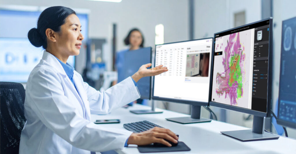
Primaa clinical validation study supported by PathPresenter

Artificial intelligence is rapidly transforming the field of pathology, but demonstrating its real-world clinical value requires carefully designed validation studies. When AI developer Primaa set out to evaluate the impact of its CLEO dermatopathology model through a clinical study, their team needed a robust platform capable of delivering whole slide images, enabling structured evaluation, and capturing detailed performance metrics from participating pathologists.

PathPresenter was proud to partner with Primaa to support this initiative, providing the secure digital infrastructure that made smooth operation of the study possible.

Evaluating the Impact of AI on Diagnostic Performance

In late 2025, Primaa began a clinical validation study to measure how its CLEO AI model could support pathologists in real-world dermatopathology diagnostic situations. The study compared diagnostic performance when pathologists reviewed cases with and without access to CLEO’s AI-powered assistance.

The goal was twofold: measure improvements in efficiency and assess gains in diagnostic effectiveness.

On the efficiency side, the study evaluated time savings per case and examined how CLEO affected specific diagnostic tasks, including:

  • Classification of lesions
  • Mitotic counting
  • Lesion measurement

For effectiveness, the study focused on key diagnostic quality metrics, including:

  • Interobserver agreement among pathologists
  • Sensitivity in detecting mitoses
  • Accuracy in classifying lesions

Cases included a range of dermatopathology diagnoses such as melanoma, nevus, squamous cell carcinoma (SCC), basal cell carcinoma (BCC), and other benign lesions, providing a realistic clinical mix for evaluation.

To conduct the study successfully, Primaa needed a platform that could provide participating pathologists with seamless access to cases, whole slide images (WSIs), and the CLEO AI model while also capturing detailed interaction data and performance metrics.

A Purpose-Built Platform enabling Clinical Studies

PathPresenter’s Image Management System (IMS) Clinical Module served as the platform for the study, with Primaa’s CLEO model seamlessly integrated into the image viewer. Given both companies’ commitment to interoperability, the integration was quickly and easily done.

Using PathPresenter, participating pathologists were able to access all study materials, including digital slides and associated case information, through a single, intuitive interface that gave easy access to the CLEO model. The platform delivered a consistent experience for each participant, ensuring standardized workflows across the study.

Equally important, the PathPresenter environment provided the security and compliance required for clinical research, meeting strict privacy and data auditability and traceability standards. This allowed Primaa to confidently share study data with participating experts while maintaining full regulatory compliance.

Beyond simple access to slides, PathPresenter enabled the structured collection of study data. The platform captured detailed metrics, including time spent per case and time required for specific diagnostic tasks, allowing the Primaa team to precisely quantify the impact of AI assistance on workflow efficiency.

Because the platform centralized all case review activity, it also made it easier for study organizers to monitor participation, ensure consistency across reviewers, and aggregate results for analysis.

Measurable Improvements in Efficiency and Diagnostic Agreement

The full results of the study are forthcoming, but in the early results from phase 1, the study has already demonstrated positive value for AI-assisted pathology workflows.

Primaa’s CLEO model enabled faster turnaround times per case, helping pathologists complete reviews more efficiently. The study also showed significant improvements in mitotic counting speed, one of the most time-consuming and tedious tasks in dermatopathology evaluation. Overall CLEO enabled a 25% reduction in analysis time.

In addition to efficiency gains, CLEO enabled a 6% improvement in diagnostic accuracy. Pathologists using the AI model demonstrated:

  • Increased interobserver agreement when classifying lesions
  • Higher sensitivity in detecting mitoses
  • Improved accuracy in lesion classification

These findings highlight the potential for AI tools like CLEO to not only accelerate pathology workflows but also enhance diagnostic reliability across practitioners.

Enabling the Future of AI Validation in Pathology

As AI continues to enter clinical practice, rigorous validation studies will play a crucial role in demonstrating safety, effectiveness, and real-world value. Platforms that enable secure collaboration, standardized workflows, and precise measurement are essential to making these studies possible.

PathPresenter is proud to support and partner with organizations like Primaa that are pushing the field forward through transparent, quantitative research.

The full results of Primaa’s CLEO validation study will be available when all phases are complete. More information about the study and Primaa’s work in AI-driven pathology, including skin and breast models, can also be found at primaalab.com.

See it in action at USCAP 2026

Primaa and PathPresenter will demonstrate their solutions at USCAP 2026 in San Antonio, March 23-25. Visit PathPresenter at booth #349 or Primaa at booth #836 for live demonstrations of the full suite of modules and integrations and more on the study results. Interested in a personal demo? Reach out to us at www.pathpresenter.com/contact

Pathology Education: The Digital Future Is Here

The next generations of pathologists are demanding digital education now

As medicine becomes more precise and data-driven, pathology is undergoing a transformative shift toward digital technology. This transition promises to revolutionize how pathologists work, improving accuracy, efficiency, and collaboration, while ultimately enhancing patient outcomes. Nowhere is this transformation happening faster or more profoundly than in pathology education. And increasingly, it is pathology trainees—residents and fellows—who are driving the demand.

A recent example from the Yale School of Medicine puts this shift into clear focus. When distinguished Yale professor and internationally recognized genitourinary pathologist Dr. Peter A. Humphrey, MD, PhD, retired in 2025, he left behind a spectacular and unique collection of pathology slides that he had used to educate generations of trainees about rare and complex cases. Over 40 years of practice, consultation, and teaching, Dr. Humphrey amassed not just slides, but insight: annotations, diagnostic pearls, and pattern-recognition wisdom refined over decades.

Through a project led by Dr. Roxanne Wadia, these slides are now being digitized (using the PathPresenter platform), along with Dr. Humphrey’s annotations, so that his experience can continue to guide new generations of residents and fellows at Yale and beyond. 

“Dr. Humphrey’s slide collection represents a lifetime of mastery, an extraordinary legacy that deserves to be more than preserved,” said Dr. Raj Singh, professor of pathology and PathPresenter co-founder. “It deserves to be shared.”

This kind of knowledge sharing is admirable, but for today’s trainees, that sharing is not just inspiring, it is becoming expected and even essential. At Yale and many other institutions, digital platforms are no longer viewed as optional enhancements. They are becoming core infrastructure for training pathologists in a world that is already digital.

The Advantages of Digital for Pathology Education

When it comes to delivering pathology education, a digital platform such as PathPresenter offers compelling benefits. But viewed through the lens of trainees, these advantages become even clearer: digital tools directly address the daily challenges residents and fellows face.

Eliminating Physical Constraints

Residents learn pathology by seeing cases—lots of them. Yet traditional training models often limit access. Glass slides are tied to specific microscopes, labs, and physical locations. Rare cases may be archived and difficult to retrieve. Multi-headed scopes allow small-group teaching, but only for those physically present.

Digital platforms eliminate these constraints. With whole-slide imaging, specimens can be digitized at high resolution and shared almost instantly with learners anywhere in the world. Instead of crowding around a microscope, trainees can review cases independently, revisit them after sign-out, and study them before board exams. They can zoom, pan, and explore at their own pace, without worrying that the slide must be returned to circulation.

For busy residents balancing clinical service, conferences, research, and exam preparation, this flexibility is transformative. Learning is no longer limited to “when the slide is available.” It becomes on-demand and continuous.

Pathology depends heavily on visual pattern recognition. Even the most detailed written description of a cellular structure cannot match the power of an image. When trainees and attendings view the exact same digital slide simultaneously, discussions are more precise. Feedback is clearer. Diagnostic reasoning becomes more transparent.

Digital images also preserve rare and valuable cases that trainees might otherwise never encounter. Instead of being stored in a single institutional archive, these collections can be shared globally. For residents preparing for subspecialty fellowships or boards, exposure to rare entities is invaluable.

This is precisely what Yale envisions for Dr. Humphrey’s collection: preserving and sharing his legacy so it can continue to guide and challenge future pathologists. Open access to world-class pathology knowledge is not just a philosophical goal, it directly supports trainee growth and confidence.

Unlocking Interactive Learning

Today’s trainees do not want to passively observe, they want to interact.

Digital platforms enable educators to annotate directly on slide images, highlight key diagnostic features, add measurements, and create guided tours through complex cases. For trainees, these annotations become enduring teaching points that can be revisited long after the live session ends.

Learners can also engage actively, leaving comments, asking questions, and even participating in real-time chats. Instead of fleeting conversations at a microscope, discussions become documented and searchable. Slide metadata, clinical history, and diagnostic commentary can travel with the image, ensuring that context is never lost.

For residents preparing for tumor boards, unknown conferences, or board examinations, this depth of context is critical. It allows them to connect morphology with clinical implications, immunohistochemistry, molecular findings, and outcomes.

Digital platforms also support structured curriculum delivery. Organized modules, case libraries, integrated quizzes, and assessments provide clarity in a training environment that can otherwise feel overwhelming. Immediate feedback helps trainees identify knowledge gaps early. Progress dashboards and certification tracking bring transparency to competency development.

In an era where pathology training varies across institutions, digital platforms help standardize access to high-quality content. For trainees, that consistency means greater confidence, regardless of where they train.

Enabling Global and Remote Learning

Trainees today are part of a global medical community, and digital pathology platforms break down geographic barriers, allowing residents and fellows to learn from experts far beyond their home institutions. A trainee in a resource-limited setting can now access the same high-quality cases and instruction as a learner at a major academic center.

Virtual tumor boards, international conferences, and cross-border case discussions are no longer rare events, they are increasingly routine. For fellows pursuing subspecialty expertise, exposure to diverse case material and global perspectives strengthens both diagnostic skill and professional networks.

For many trainees, remote learning is not a compromise; it is an opportunity, and perhaps even a necessity.

Integrating Artificial Intelligence into Training

The pathologists of tomorrow will practice in environments shaped by artificial intelligence. AI models are already being integrated into clinical workflows and research applications, from image analysis to diagnostic support.

Trainees recognize this shift. They want to understand not only how to interpret slides, but how to evaluate and leverage AI tools responsibly.

Comprehensive digital platforms provide a natural environment for this integration. In a digital workspace, residents can explore AI-assisted analyses, compare algorithmic outputs with their own interpretations, and develop the critical thinking skills necessary to use these tools wisely.

Exposure during training ensures that future pathologists are fluent in both traditional morphology and digital augmentation. Digital literacy is becoming as fundamental as microscopic technique.

Meeting Trainee Expectations

Today’s residents and fellows are digital natives. They expect intuitive interfaces, instant access to resources, and collaborative tools that resemble the platforms they use in other aspects of their lives.

They also understand that their future employers (academic centers, hospitals, laboratories) are moving toward digital pathology workflows. They want their training environment to reflect that reality.

Institutions that embrace digital platforms signal to trainees that they are forward-looking and invested in preparing them for modern practice. Those that do not risk falling behind, not only technologically, but competitively in recruiting top talent.

The Digital Future Is Now

The shift toward digital pathology education is already underway, and trainees are at its center. The benefits are clear: expanded access, interactive learning, global collaboration, standardized curricula, AI integration, and preparation for real-world practice.

As the practice of pathology becomes increasingly digital, education must mirror that evolution. For residents and fellows, digital platforms are not optional enhancements. They are essential tools for mastering the discipline.

Let Us Help You on Your Path

PathPresenter was created specifically to unlock the opportunities that a digital education platform offers. With tens of thousands of users, our public platform is the world’s largest pathology education community. For institutions, our enterprise education module enables organizations to manage, deliver, and generate measurable ROI on digital pathology education, while integrating seamlessly with clinical practice and research.

Whether you are modernizing your residency curriculum, digitizing legacy collections, hosting global conferences, or building a private learning portal for trainees, PathPresenter has a powerful platform to help you. We would be delighted to discuss your pathology education goals.

The next generation of pathologists is ready for a digital future. Let us help you build it.

Why a Vendor-Agnostic IMS Is Key to a Future-Proof AI Strategy

by Patrick Myles, MBA
CEO, PathPresenter

As AI adoption accelerates in digital pathology, many institutions are facing a strategic decision that will shape their technology stack for years to come: how tightly should AI be coupled to the core imaging and workflow platform?

At PathPresenter, we’ve taken a very deliberate position. On the AI side, we are intentionally AI-agnostic at the IMS and workflow level. That decision is rooted not in theory, but in hard-earned lessons from the field.

The Hidden Cost of Closed AI Ecosystems

Over the years, we’ve seen multiple institutions make large investments in closed scanning and workflow ecosystems, often driven by early regulatory approvals or the promise of an “end-to-end” solution. In the short term, those systems worked. But over time, they frequently became bottlenecks.

When institutions later wanted to onboard additional scanners, integrate third-party tools, or adopt stronger algorithms from competing vendors, those closed platforms made change slow, expensive, or in some cases impossible. The result was delayed deployments, integration failures, and ultimately costly replacement projects that disrupted operations far more than anyone anticipated.

That model simply doesn’t hold up in today’s AI landscape.

AI Is Changing Too Quickly for Locked-In Platforms

AI is moving at a pace we’ve never seen before. Many algorithms deployed in production today are CNN-based, while the next wave of capability is clearly shifting toward foundation-model approaches. As performance improves and new vendors emerge, the “best” solution will continue to change.

In that environment, locking the core IMS and workflow layer to a specific AI vendor is a strategic risk. Our view is simple: the IMS should remain independent of the AI layer.

A Future-Proof Strategy that Keeps the Platform Stable While AI Evolves

To make that separation practical, we’ve invested heavily in building an AI middleware layer designed to be future-proof. This middleware can ingest, normalize, and orchestrate the clinical and operational data required to deploy AI at scale, including integrations built on HL7, FHIR, and DICOMweb. By abstracting these complexities away from the core platform, institutions can adopt new AI models and workflows over time without needing to rebuild integrations or re-platform each time the ecosystem evolves.

Just as importantly, this approach is informed by real-world experience. We’ve worked with a wide range of institutions and organizations and have seen multiple generations of digital pathology and AI vendors come and go. Some early companies were highly innovative, but struggled to scale or keep pace with market shifts.

Those experiences have consistently reinforced the same conclusion: workflow platforms must remain stable, while the AI layer must remain flexible.

In an AI landscape defined by constant change, future-proofing isn’t about predicting which algorithm will win. It’s about designing systems that can evolve, without disruption, as the technology does.

About the Author

Patrick Myles is CEO of PathPresenter. Previously, he was CEO of Huron Digital Pathology, and Vice President of Business Development for Teledyne DALSA. He served as a board member of the Digital Pathology Association.

The ROI of Remote Consultations

Remote second opinions can be a gateway to proving the value of digital pathology

The ROI of Remote Consultations in Digital Pathology

Digital pathology has reached an important inflection point. Adoption is rising, evidence of clinical and operational value is strong, and digitization is widely accepted as the future of pathology. Yet as Imogen Fitt of Signify Research pointed out in her recent analysis of the market, near-term adoption remains uneven. For many institutions, digital pathology still feels like something to plan for tomorrow rather than act on today.

The reasons are well understood. High upfront costs, complex and lengthy implementations, interoperability challenges and heavy administrative overhead all combine to slow decision-making. Even organisations that are philosophically aligned with digital pathology often struggle to justify investment when budgets are under pressure from other enterprise-scale initiatives such as LIS replacements, oncology platforms or radiology IT refreshes. The result is a persistent gap between recognised long-term value and near-term return on investment.

What many organizations are seeking is not another abstract promise about future AI or biomarker discovery, but a way to realise tangible benefits quickly, in a form that aligns with their specific clinical and operational realities. This is where a more targeted, use-case-driven approach to digital pathology can make a meaningful difference.

Thinking outside the building

For institutions with significant subspecialty expertise, remote second opinion services represent one of the most compelling and immediately actionable applications of digital pathology. They address real clinical needs today: managing rising case complexity, improving access to scarce expertise (just locally but even globally), supporting workforce sustainability and generating new, scalable revenue streams. PathPresenter’s ConsultConnect was designed specifically to unlock this opportunity while removing many of the barriers that slow broader digital pathology adoption.

ConsultConnect is a secure, “zero-footprint” cloud-based platform built to enable remote pathology consultation at scale, allowing consulting institutions with specialty expertise to take referral cases from remote organizations with ease. Rather than requiring a full digital pathology rollout, it allows organisations to start with a focused, high-value workflow that delivers rapid impact. This fundamentally changes the cost–value equation.

First, ConsultConnect eliminates the issue of high upfront costs. There is no capital expenditure on IT infrastructure and partners such as Grundium can provide pathways to cost-efficient slide scanners optimized for rapid deployment. Instead of large initial outlay and a long procurement cycle, the ConsultConnect model is based on a minimal annual commitment with simple, transparent “per-consult” pricing. This makes financial planning easier and aligns cost directly with value generated.

Second, it removes the burden of complex implementations. Traditional DP projects can take months or years to move from approval to production, often involving shared IT infrastructure, local software installation and extensive validation. ConsultConnect deploys in days, not months. Because it is “zero-footprint” cloud-native and requires no shared infrastructure between organisations, it avoids many of the technical bottlenecks that stall progress.

Interoperability is another major friction point in digital pathology adoption, particularly as laboratories seek to simplify fragmented IT environments. ConsultConnect removes this barrier by accepting images from virtually any whole slide scanner or file format. Institutions do not need to standardise on a single vendor or invest in expensive integration work to participate, making it an ideal solution in heterogeneous or transitional environments.

Administrative hurdles will always vary by organization, but ConsultConnect significantly reduces their impact by minimising IT, funding, and operational complexity. Importantly, it also offers something many DP initiatives lack: a fast, credible path to revenue generation. By enabling institutions to scale remote second opinion services securely and efficiently, the platform turns digital pathology from a cost centre into a potential revenue driver.

The result is a low-risk, high-impact entry point into digital pathology. Institutions can support referring pathologists, extend the reach of their subspecialty expertise, and deliver measurable ROI in the near term, without waiting for large-scale transformation programs to clear internal hurdles.

From “Why Now?” to “Yes, Now”

In a market where “why now?” remains one of the hardest questions to answer, ConsultConnect provides a practical response. It demonstrates that digital pathology does not have to mean extra work, excessive risk or deferred value. Instead, it can be a rational, incremental step that delivers tangible benefits today while laying the foundation for broader digital adoption tomorrow.

Does your institution have expertise to share, ambitions to scale, and aspirations for digital transformation? ConsultConnect could be right for you. We’d love to discuss your needs.

Behind the Scenes of a Successful FDA 510(k): PathPresenter’s Clinical Viewer Journey

by Brian Matcheski
Regulatory Manager, PathPresenter

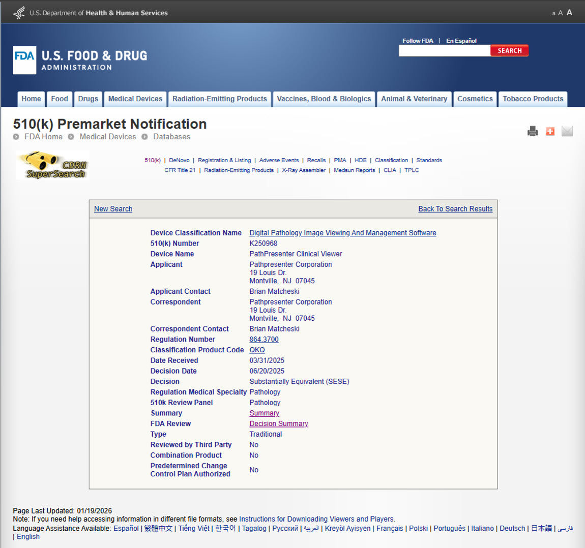
PathPresenter's FDA 510(k) Premarket Notification

FDA 510(k) clearance is often described as a milestone in digital pathology. What’s discussed less is how you actually get there, especially for software-only image viewers, where regulatory expectations differ meaningfully from scanners and other hardware-based systems.

With the recent FDA 510(k) clearance of the PathPresenter Clinical Viewer, we wanted to share some perspective on our journey: what mattered most, what surprised us, and why a disciplined approach to the process ultimately pays dividends. This isn’t a detailed how-to guide, and there are no shortcut hacks to share (with the possible exception of “get good advice from a qualified regulatory expert with experience in digital pathology”). But for those involved in navigating FDA clearance for digital pathology software, these insights from our journey may prove interesting.

Why FDA Clearance Matters in Digital Pathology

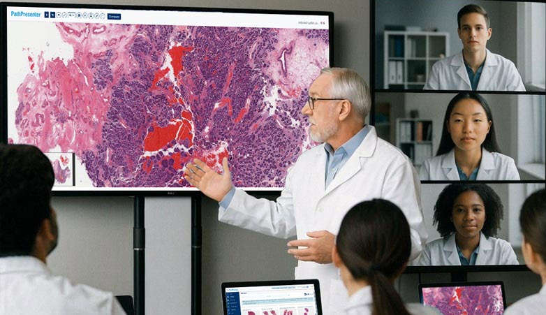
Review meeting

As digital workflows in pathology move from research into routine clinical use, regulatory oversight becomes essential to building trust and enabling adoption. FDA regulation of digital pathology software ensures that clinical tools meet standards for safety, effectiveness, and reliability. 

The FDA has several pathways for market clearances, but to date, digital pathology devices have concentrated on just two: de novo and 510(k). The de novo (Latin for “new”) pathway is used for new devices with no existing precedent (or predicate). The 510(k) pathway applies when a new device can demonstrate substantial equivalence to an existing, legally marketed predicate. Clearly, the 510(k) pathway is only available when a predicate already exists; it clears a new component to do an equivalent job in an existing system. Not surprisingly, de novo clearances typically involve greater regulatory uncertainty, broader risk justification, and longer timelines than 510(k). 

Neither clearance pathway represents FDA “approval,” nor do they validate clinical outcomes, but clearance is a critical prerequisite for deploying software in regulated diagnostic environments.

For pathologists and labs, FDA clearance provides confidence that a viewer can be used in primary diagnostic workflows. For healthcare systems and enterprise customers, it reduces risk and enables broader clinical integration. For PathPresenter customers, our 510(k) clearance unlocks the ability to deploy our Clinical Viewer in regulated clinical settings with confidence.

Understanding Classification, Intended Use, and Predicates

One of the earliest and most important steps in the process for PathPresenter was clearly defining intended use. Digital pathology viewers are regulated differently from whole slide imaging (WSI) scanners. While scanners capture the image, viewers render, display, and enable interpretation of that image, and the FDA evaluates them separately.

That distinction drives selection of the appropriate predicate device. Substantial equivalence hinges not only on features, but on intended use, performance characteristics, and risk profile. Getting alignment here early set the foundation for the entire submission.

Planning to Succeed: Following the FDA’s Guidance

One of the best decisions we made was to anchor our submission directly to FDA guidance documents, including:

  • Technical Performance Assessment of Digital Pathology Whole Slide Imaging (WSI) Devices
  • Content of Premarket Submissions for Device Software Functions
  • Cybersecurity in Medical Devices: Quality System Considerations and Content of Premarket Submissions

These documents do more than list requirements, they provide a window into how FDA reviewers think about risk, safety, and effectiveness. By structuring our submission around this guidance, we weren’t guessing what the FDA wanted to see. We were aligning with how the agency approaches review.

A strong 510(k) submission is not about volume. It’s about completeness, traceability, and clarity.

Working with FDA Reviewers

Going into the review, we were cautious. In mid-2025, widely reported austerity measures affecting HHS and the FDA raised concerns across the industry about review timelines and resource constraints.

In our experience, those concerns did not materialize. The FDA’s feedback throughout the review was timely, thoughtful, and constructive. Importantly, our submission was reviewed and cleared within the FDA’s stated 90-day service level.

That experience reinforced our confidence in both the process and the people behind it. Even in a challenging operating environment, the review team remained focused on scientific rigor, patient safety, and predictable execution.

Investing Early Where It Mattered Most: Pixel-Wise Image Comparison

For software-only digital pathology viewers, one of the most scrutinized aspects of a 510(k) submission is pixel-wise image comparison to the predicate viewer. This is an area where other OEMs have historically faced delays.

For a clinical viewer, the primary safety risk is not image acquisition but image presentation. Even small rendering differences, such as altered color balance, contrast, or interpolation, could theoretically influence a pathologist’s interpretation. Pixel-wise comparison evaluates whether the same whole slide image (WSI), when rendered by two different viewers, produces image outputs that are effectively the same at the pixel level. The goal is to show that the subject viewer does not introduce image artifacts, distortions, or rendering differences that could impact clinical interpretation.

We anticipated scrutiny in this area and invested significant effort upfront. That meant carefully defining our comparison methodology, validating our approach, and presenting results that were clear, reproducible, and defensible.

That investment paid off. When FDA reviewers conducted their own image evaluations, they were able to quickly confirm that our viewer met performance expectations, without prolonged back-and-forth. It was one of the most valuable early investments we made in the process.

Responding Quickly and Completely to FDA Feedback

Even with a thorough submission, FDA reviewers are trained to identify areas of potential risk or ambiguity (and they should). We treated every FDA comment as an opportunity to strengthen the submission.

We made our responses timely, direct, and supported by data. There are no shortcuts here. That responsiveness helped maintain momentum and avoid unnecessary review cycles, which can otherwise add weeks or months to the process.

Treating Pre-Submission Feedback as a Contract

Part of the submission process includes Pre-Submission (Pre-Sub) meetings with the FDA. These can be very valuable, but only if the feedback is taken seriously. We treated Pre-Sub input as a contract, not a suggestion.

Every item raised during those discussions was addressed explicitly, point by point, in the final submission. If you’re not prepared to incorporate or directly respond to Pre-Sub feedback, it’s fair to ask why you’re having the meeting at all. Alignment early pays off later.

Building Clinical Trust Through Regulatory Excellence

For PathPresenter, FDA 510(k) clearance is not just a regulatory checkbox. It reflects our commitment to building clinical software that pathologists can trust in real-world diagnostic workflows.

We’re proud of the clearance, and equally proud of the disciplined, transparent approach that got us there. As digital pathology continues to evolve, we remain committed to quality, safety, and innovation, grounded in a regulatory process that exists for a reason.

If you’d like to learn more about the PathPresenter Clinical Viewer or see it in action, we invite you to request a demo or reach out to our team.

The Digital Pathology Illusion: Why Are We Building Storage Instead of Solutions?

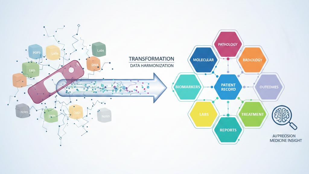
Transformation and data harmonization

Digital pathology has finally moved from “nice-to-have” to a global strategic priority. The benefits are real and include remote sign-out, operational efficiency, collaboration, education, and the promise of AI. But as health systems rush to digitize, many are falling for a dangerous misconception: that digital slides automatically equal a valuable dataset.

They don’t.

Right now, we’re witnessing a massive and expensive race to digitize pathology, yet we’re often solving the wrong problem. If your organization’s primary focus is “where to store the pixels,” you’re not building a future for AI, on the contrary, you’re building a more expensive filing cabinet.

The truth: no one wants your slides

It’s time for a reality check: no researcher, pharma partner, or AI developer is looking for “just digital slides.” A WSI in isolation is a clinical dead end and actually a financial liability. Every day that pixel data sits in a silo without clinical context, it consumes storage costs while generating zero insights. A slide only becomes valuable when it’s connected to the patient’s clinical truth and metadata that link to tumor type and subtype, molecular and prognostic markers, treatment course, and outcomes (response, recurrence/progression, survival). Without that context, a WSI is often just a beautiful high-resolution image file with limited real-world utility.

We digitized the workflow, not the intelligence.

Most institutions today have a “digital pathology” environment that is still a graveyard of silos: slides live in scanner storage or cloud buckets, reports sit elsewhere in the LIS/EMR as static PDFs, and molecular results, labs, treatments, and outcomes remain buried in separate systems or unstructured clinical notes. Even within pathology, the report is still largely narrative text, so downstream “metadata” often stops at age, sex, and diagnosis, while key details like biomarkers, margins, tumor size, staging, and prognostic factors remain trapped in free text. In short: we’ve digitized pathology, but we haven’t structured it, and without structure, we don’t have a dataset, we have digital clutter.

The real dataset is a unified patient story

Real value of the kind that accelerates research, supports precision medicine, and enables reliable AI only exists when data is multi-modal, longitudinal, and semantically organized. That means slides are programmatically linked to pathology findings, biomarkers/molecular data, labs, radiology, treatment events, and outcomes, following the patient journey over time. Researchers don’t search for “tumor” vs “cancer” vs “malignancy”. They search by clinical meaning: triple-negative breast cancer, EGFR-mutated lung adenocarcinoma on targeted therapy, high-risk disease with recurrence. That requires semantic organization, not just storage.

And this is becoming urgent. We’re entering the era of tabular foundation models, where AI learns from structured clinical tables at scale. These models are only as good as the structure and consistency of the data they ingest. Messy, disconnected silos don’t just slow AI down, they undermine its reliability.

The bottom line

“The goldmine of pathology data” will remain a hollow promise until we stop treating the image as the final product. The image is the starting line. The dataset is the unified, searchable, longitudinal record of the patient’s life.

Stop asking: “Where do we store the slides?” Start asking: “How do we build a structured data foundation that connects the pixels to the patient, automatically and at scale?”

If you aren’t solving for data structure, you aren’t solving the problem.

Pathologists are not just the users of these systems; they are the architects of the clinical truth. If the pathologist doesn’t define the structure at the point of capture, the data is lost to the “narrative abyss” of a PDF forever. But realizing its full translational value requires partnership with strong technical teams who understand structured data design, concept libraries and standardized ontologies, flexible database architectures, tabular foundation models, and semantic search. Together, we can turn stored pixels into the most valuable data in healthcare that powers trustworthy AI and ensures every patient receives the right treatment at the right time.

About the Author

Dr. Rajendra Singh is a Professor of Pathology at the University of Pennsylvania and co-founder of PathPresenter. He serves as a member of the Digital and Computational Pathology Committee of the CAP, Editorial Board of the WHO for Classification of tumors, 5th Edition and the Board of Digital Pathology Association.

Success Story: Accelerating Clinical Trials for Oncology with Imagenomix Predict and PathPresenter ConsultConnect

Zero-footprint web portals and AI-enhanced workflows reduce screening bottlenecks for an AKT-Pathway clinical trial

Laboratory scientists

In Brief

  • Oncology research trials require patients but enrollment can be a major limiter. 40% of oncology trials fail due to under-enrollment and 80% do not meet enrollment timelines
  • Imagenomix addresses the enrollment challenge: Imagenomix’s Predict AI model for rapid mutation prediction from H&E slides speeds patient enrollment in clinical trials and lowers trial costs
  • To recruit enough patients, clinical trials often include dozens of trial sites. This creates new challenges in collecting and aggregating data for analysis and reporting when those sites do not share the same laboratory and IT systems
  • PathPresenter ConsultConnect solves the interoperability challenge by accepting and aggregating data from all trial sites into one data store, allowing efficient, concentrated and centralized analysis and reporting. Being cloud based, vendor-agnostic and built for interoperability, ConsultConnect requires no shared IT infrastructure or local software installation, and it accepts images from virtually any whole slide scanner or format.

The Challenge of Patient Enrollment

Clinical trials are the lifeblood of oncology research, providing the evidence that transforms promising therapies into approved treatments. Yet, patient enrollment remains one of the most persistent obstacles to trial success. Studies show that 40% of oncology trials fail due to under-enrollment, and 80% fail to meet enrollment timelines. The consequences are significant: delays in bringing lifesaving therapies to patients, higher trial costs, and incomplete or inconclusive data.

A key reason for insufficient enrollment lies in the complexity of matching the right patients to the right trial in timely fashion. Molecularly driven oncology trials depend on identifying specific genetic mutations, gene fusions, or molecular signatures in a patient’s tumor. These molecular biomarkers determine eligibility and help researchers understand which patients are most likely to benefit from an investigational therapy. Unfortunately, traditional laboratory workflows are slow, labor-intensive and expensive. Next-generation sequencing (NGS), while accurate, typically takes three to four weeks to deliver results and requires substantial tissue samples. The cost and delay not only slow patient enrollment but may also disqualify patients whose clinical status changes before testing is complete.

Accelerating Enrollment with AI

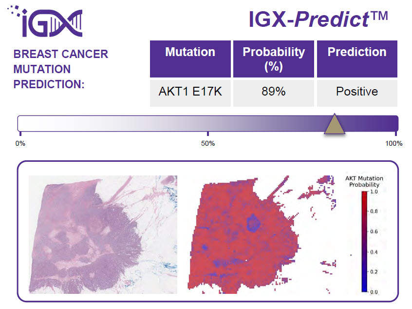
Imagenomix set out to solve this enrollment challenge by developing a faster, more scalable approach to patient screening through digital pathology and AI predictive models. Its IGX Predict AI-based technology analyzes the H&E-stained biopsy whole slide images that are already part of digital pathology workflows to predict the presence of key genetic mutations. Using advanced deep-learning models, IGX Predict identifies subtle histologic patterns correlated with genomic alterations, returning results in minutes instead of weeks. Predicted mutations are then confirmed by a multi-gene panel or a low-cost single gene assay, enabling clinical trial screening and enrollment even in low-income settings, for patients who do not have access to large NGS panels.

IGX Predict

IGX Predict AI mutational screening enables clinicians and researchers to assess far more patients, more quickly, and at lower cost. Instead of waiting weeks for sequencing results, trial coordinators can rapidly identify patients who are likely to qualify for a given study and prioritize them for confirmatory testing or immediate enrollment. The result is faster trial startup, reduced screening costs, and greater access for patients who may benefit from investigational therapies.

By using AI to triage patients and predict mutation profiles, Imagenomix helps sponsors overcome one of the most critical barriers in oncology research: finding enough qualified participants to power statistically meaningful trials in a timely fashion.

From Enrollment Success to Data Management Complexity

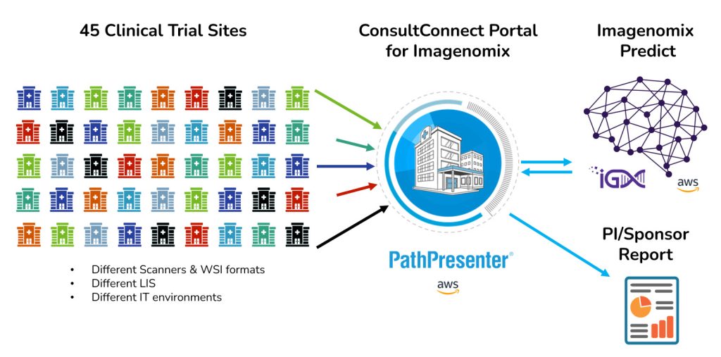
In a current multi-site oncology study, the Imagenomix approach is enabling rapid enrollment across 45 clinical trial centers. With the enrollment hurdle cleared, a new challenge emerges: how to manage and harmonize the flood of data generated by dozens of institutions operating with different laboratory and IT infrastructures.

Each center has its own scanners, laboratory information systems (LIS), and compliance requirements. Some hospitals prohibit local software installation, making it difficult to deploy analysis tools or data transfer agents. Trial sponsors and investigators need a single, secure system to collect, store, and analyze digital pathology data from every site, regardless of scanner type, file format, or local IT policy.

Consistent, centralized access is also essential for quality control and reporting. Pharmaceutical sponsors require unified access to trial results and the ability to review data from all sites without the delays caused by manual data consolidation.

The Solution: PathPresenter ConsultConnect

To address these challenges, Imagenomix partnered with PathPresenter, integrating its ConsultConnect platform as the data backbone of the trial. Designed for multi-site, multi-platform interoperability, ConsultConnect is a cloud-based, vendor-agnostic solution that accepts images from virtually any whole-slide scanner or file format. It requires no local installation or shared IT infrastructure, making it ideal for diverse hospital environments with strict security policies.

Within days, PathPresenter deployed a secure ConsultConnect web portal on AWS specifically for the Imagenomix Predict platform. Using this platform, each pathology laboratory and trial site can safely upload data directly to the portal without any software installation, slide image adjustments or configuration burden. Each site sees only their respective cases while participating in a large multicenter study. The platform’s intuitive web interface built for pathologist by pathologists requires minimal training and facilitates rapid adoption across geographically distributed teams. Each site and each submitting pathologist has unique login credentials, providing traceability and ensuring no data leakage between sites.

ConsultConnect Portal for Imagenomix clinical trial

Once uploaded, the data is automatically organized into a centralized repository. Imagenomix’s IGX Predict AI algorithms operate in the cloud to analyze the slides and generate mutation predictions. Results are seamlessly delivered back through the ConsultConnect portal, where they can be accessed by trial investigators and sponsors.

ConsultConnect’s robust healthcare-grade security and privacy controls ensure compliance with patient data protection regulations, while its scalable cloud architecture supports efficient, high-volume processing of digital slides.

Results and Impact

The combination of Imagenomix Predict and PathPresenter ConsultConnect is transforming the operational efficiency of the trial.

  • Faster Trial Execution: With IGX Predict’s AI-based mutation prediction, patient identification that once took weeks now takes minutes, accelerating recruitment and trial initiation.
  • Simplified Data Management: ConsultConnect eliminates the need for complex software deployments and manual data aggregation. All centers are able to contribute data seamlessly to a unified cloud environment.
  • Enhanced Collaboration: Investigators and sponsors gain centralized visibility into results, supporting real-time collaboration and decision-making across sites.
  • Improved Cost and Efficiency: Reduced reliance on costly molecular testing and streamlined data workflows translates into substantial savings in time, tissue and cost.
  • Scalable Model for Future Trials: The success of this implementation establishes a repeatable, scalable model for future oncology and biomarker-driven studies involving multi-site data collection.

A Blueprint for the Future of Oncology Research

This case study illustrates how combining AI-driven mutation prediction with cloud-based interoperability can redefine the pace and efficiency of clinical research. Imagenomix Predict tackles the most pressing barrier of patient enrollment, while PathPresenter ConsultConnect resolves the data-logistics challenge inherent in large, distributed trials.

Together, they enable sponsors to run faster, more cost-effective studies without compromising scientific rigor or patient privacy. As oncology trials continue to grow in complexity and precision, such integrated, interoperable technologies will be indispensable in bringing the next generation of targeted therapies to patients worldwide.

More Information

Interested in learning more about how these solutions can help solve your own challenges? Contact us.

Why Seamless IMS-LIS Integration Is Essential for Digital Pathology

Unified digital workflows connecting IMS to LIS unlock efficiency, compliance, and the full potential of AI for modern pathology

Digital pathology has moved decisively from experimentation into everyday practice. For pathology organizations, it now underpins clinical workflows, operational efficiency, and long-term strategy. To enable remote sign-out and AI-driven analysis, organizations have turned to Image Management Systems (IMS) that can organize, track, and contextualize whole slide images across the diagnostic workflow.

However, the data needed to interpret those images extends well beyond what an IMS typically manages. Patient demographics, clinical context, specimen metadata, test orders, results, finalized reports and more all live in a separate but equally critical system: the Laboratory Information System (LIS). For groups accustomed to thinking primarily in terms of image workflows, the LIS can feel like a parallel universe, yet it is the system of record for nearly all non-image pathology data. When IMS and LIS platforms operate in isolation, inefficiencies emerge, context is lost, and the promise of digital pathology remains only partially realized.

As digital pathology programs mature, it is becoming evident that success depends not only on performance, scale, and regulatory readiness, but also on seamless interoperability. In this article, we explore why deep, context-aware integration between an IMS and a modern LIS is essential to unlocking the full value of digital pathology. We’ll examine the practical challenges this integration resolves and the strategic benefits it enables for pathology groups advancing toward a fully digital, AI-enabled future.

Image Management Systems (IMS) in Digital Pathology

A digital pathology image management system (IMS) goes far beyond mere image viewing. At its best, it is an enterprise-grade platform designed to manage the entire lifecycle of digital pathology images across clinical, research, and educational workflows, and serving as the backbone of digital pathology operations, supporting everything from secure storage to AI integration and multi-institution collaboration.

Core Capabilities of a Modern Digital Pathology IMS

A robust IMS typically includes:

  • Advanced Slide Viewing: High-performance visualization across multiple formats (SVS, NDPI, SCN, DICOM, BigTIFF, and more), synchronized multi-slide viewing, annotations, overlays, and fast rendering.
  • Centralized Data Management: Secure, scalable image storage, on-premises, cloud, or hybrid, with governance controls and long-term data stewardship.
  • Security and Compliance: HIPAA-grade protections, encryption, audit trails, role-based access, and regulatory readiness for clinical use.
  • Workflow Integration: Case management, task tracking, and seamless interoperability with LIS, EHR, PACS, and other enterprise laboratory software systems.
  • User Roles and Access Control: Granular permissions ensure that only authorized users access sensitive data, while maintaining accountability.
  • Collaboration Tools: Support for remote consults, tumor boards, education, and peer review through shared environments.
  • AI and Image Analysis Enablement: Integration with AI models for detection, quantification, and pattern recognition, embedded directly into diagnostic workflows.
  • Regulatory Readiness: Many IMS platforms are designed to meet FDA, HIPAA, GDPR, IVDR, and other regulatory standards. Some, such as PathPresenter’s clinical viewer, are FDA 510(k) cleared and EU-IVDR certified for primary diagnosis when paired with approved scanners.

In short, an IMS provides a comprehensive infrastructure for digital pathology. It can be highly capable on its own, handling slide storage, visualization, annotations, and even AI outputs. But when it operates independently from the laboratory’s core operational system, its impact is inherently limited. Rather than dissolving legacy barriers, a disconnected IMS can unintentionally recreate them in digital form.

True digital pathology maturity is reached only when images and laboratory data function as a single, coherent workflow. Without that alignment, pathology groups often find themselves managing parallel systems that never quite converge.

The Operational Cost of Keeping IMS and LIS Separate

When image workflows and laboratory workflows are not connected at a foundational level, pathology teams encounter familiar but amplified challenges, including:

  • Reliance on manual reconciliation of cases and slides
  • Repeated entry of patient and specimen data
  • Frequent toggling between systems during case review
  • Greater exposure to mismatches, omissions, and reporting errors
  • Disjointed compliance records spanning multiple platforms
  • Collaboration slowdowns across sites and subspecialties

In isolation, each issue may seem manageable. At scale however, particularly in regulated, high-throughput environments, they compound rapidly, eroding both efficiency and confidence in the digital workflow.

Rethinking the LIS: More Than a Back-End Database

For many digital pathology teams, the LIS is perceived primarily as a static repository: the place where orders originate and reports ultimately land. While that historical role remains essential, it no longer reflects how modern laboratories operate.

From Passive Record-Keeping to Active Workflow Orchestration

Today’s laboratories increasingly require systems that do more than store information. A modern LIS such as LigoLab functions as an operational engine: driving workflow logic, enforcing rules, automating handoffs, and presenting the right context at the right moment.

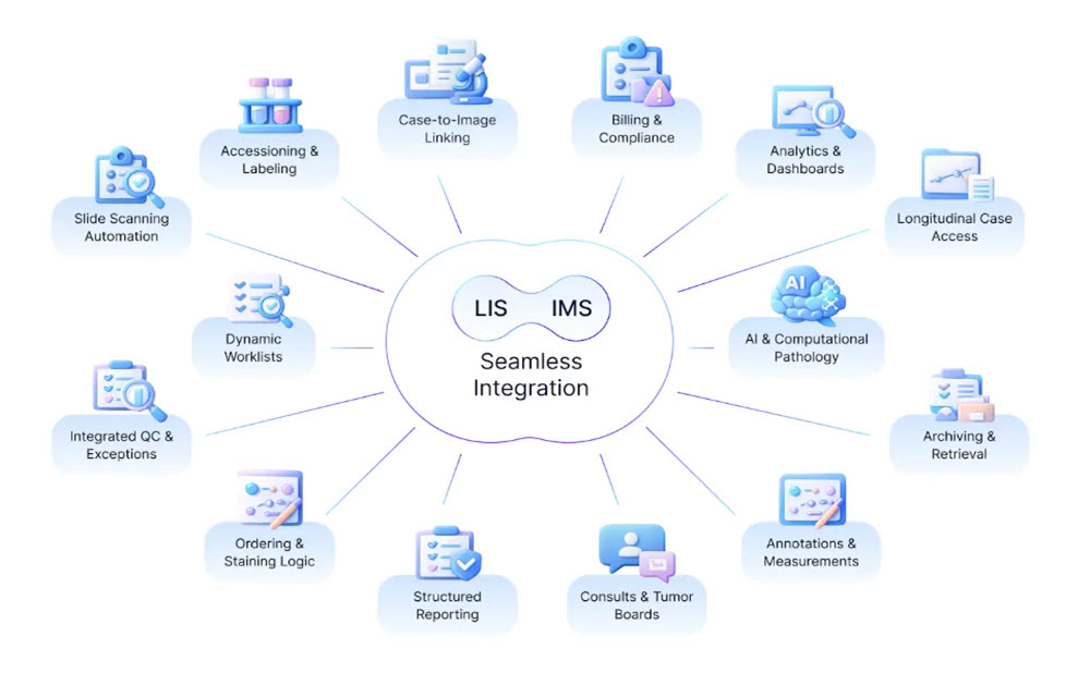
When an IMS is natively integrated into this environment, digital pathology is no longer an external tool that users “visit.” Instead, image review becomes a natural extension of the laboratory workflow, governed by the same logic, permissions, and traceability as the rest of the case lifecycle.

What Integrated LIS–IMS Workflows Enable Day to Day

In a genuinely unified digital pathology environment:

  • Digital slides are opened directly from the laboratory case, not searched for separately
  • Patient, specimen, and order context flows automatically into image review
  • Movement between data, images, and reports feels continuous rather than segmented
  • Auditability covers both diagnostic decisions and image interactions end to end
  • AI outputs appear within established workflows rather than as side-channel tools

This level of integration changes how work is performed: not by adding features, but by removing friction.

LIS-IMS integration

Core Challenges Solved by Deep Integration

1. Smoother, Faster Diagnostic Workflows

When image access is embedded within the laboratory system, pathologists spend less time navigating software and more time interpreting cases. Reduced friction translates directly into faster turnaround and lower cognitive load.

2. Scalable Support for Remote Practice

Integrated platforms make location largely irrelevant. Secure remote sign-out, real-time collaboration, and distributed subspecialty review become standard operations rather than exceptions.

3. Better Decisions Through Complete Context

Images rarely tell the full story on their own. Tight coupling between LIS and IMS ensures that diagnostic interpretation always occurs alongside the complete clinical and specimen context, reducing the risk of incomplete assessment.

4. Stronger Governance and Compliance

When images and laboratory data share a unified audit framework, compliance with regulatory and accreditation requirements becomes simpler and more defensible—without added administrative burden.

5. A Practical Path to Enterprise AI

AI delivers value only when it operates inside real workflows. Integrated LIS–IMS environments provide the governance, data access, and workflow hooks required to deploy AI responsibly and at scale.

Integration in the Real World: From Concept to Practice

A practical example of this approach can be seen in the collaboration between PathPresenter and LigoLab. Rather than treating image management as an external system, PathPresenter’s clinical viewer is embedded directly within LigoLab’s enterprise laboratory platform.

The result is a unified diagnostic experience where pathologists interact with slides in the same environment where cases are managed, orders are tracked, and reports are finalized. This tight coupling reduces navigation overhead, accelerates review cycles, and establishes a solid foundation for future AI-driven enhancements.

Related: LigoLab and PathPresenter Announce Strategic Partnership to Deliver Seamless Digital Pathology Workflows

Building Toward a Unified Digital Pathology Future

At its core, LIS–IMS integration is not about software convenience. It is about redefining how pathology work flows from accessioning through diagnosis and reporting.

When laboratory data and images operate as one system:

  • Operations gain visibility and automation
  • Financial teams benefit from cleaner alignment with billing and revenue workflows
  • Clinical staff gain modern tools that reduce manual effort
  • Patients benefit from faster, more consistent diagnostic outcomes

As digital pathology, AI, and remote diagnostics continue to evolve, this alignment moves from “nice to have” to mission critical. Pathology groups that invest early in deep, contextual integration position themselves not just to digitize slides, but to fundamentally modernize how pathology is practiced.

More Information

Looking for a new (or improved) LIS-IMS integration? Connect with our team to assess your workflow needs and explore the best solution.

Digital Pathology Basics: Image Viewers vs. Image Management Systems

Image Viewer vs IMS comparison

In brief

  • An image viewer focuses on the basics of viewing and interacting with digital slides.
  • A digital pathology IMS manages the entire lifecycle of digital pathology images within a clinical, research, or educational workflow.

As digital pathology becomes more integrated into clinical, academic, and research settings, it’s important to distinguish between the tools used in this domain. Two common types of software encountered are pathology slide viewers and digital pathology image management systems (IMS). While they may appear similar on the surface (both enable users to access and interact with digital slides) they are fundamentally different in terms of scope, features, and intended use.

Pathology Slide Viewer: A Focused Viewing Tool

A pathology slide viewer is a software application designed primarily for visualizing digitized pathology slides, also known as whole slide images (WSIs). These tools typically allow users to open and navigate high-resolution scans of tissue specimens. Common features include zooming, panning, rotation, and the ability to add simple annotations or measurements.

These viewers can be used by pathologists for reviewing cases, by students for learning, or by researchers for image analysis. They may be standalone desktop applications or web-based platforms. Some viewers are vendor-specific, tied to a particular scanner or image format, while others are open source or multi-format compatible. However, pathology slide viewers are limited in scope. Their main purpose is to enable manual, visual inspection of slides. They typically do not provide broader workflow functionality, user management, or system integrations. For small-scale or individual use, slide viewers are handy tools. But their limitations will quickly become apparent.

Digital Pathology Image Management System (IMS): A Comprehensive Platform

In contrast, a digital pathology image management system (IMS) such as PathPresenter is a comprehensive, enterprise-grade software solution that manages the entire lifecycle of digital pathology data. It includes not only the viewing of whole slide images, but also the organization, tracking, analysis, and sharing of those images in clinical and research workflows. An enterprise deployment of an integrated digital pathology workflow will almost certainly require a fully featured IMS.

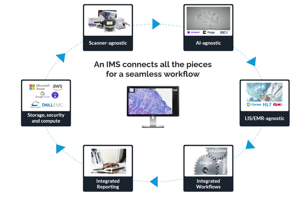
IMS connects all the pieces for a seamless workflow

A robust IMS typically includes the following core capabilities:

  • Slide Viewing: IMS platforms provide high-quality visualization with features like synchronized multi-slide viewing, annotation, overlays, and fast rendering across many file formats (svs, ndpi, scn, dcm, bigtiff, etc.).
  • Data Management: IMS centralizes and secures pathology data with on-premise, cloud, or hybrid storage options.
  • Security: IMS must meet strict standards (e.g., HIPAA), ensuring PHI confidentiality, integrity, and availability while protecting against threats.
  • Workflow Integration: Platforms support case management, task tracking, and diagnostic, research, or education workflows, often with integrated security like SSO.
  • User Roles & Access Control: Permission-based access and audit trails ensure only authorized users can view sensitive data while maintaining accountability.
  • Interoperability: IMS integrates with LIS, PACS, EHR, and other systems. PathPresenter is vendor-agnostic, with integrations to leading systems such as EPIC Beaker and LigoLab.
  • Collaboration Tools: Multi-user environments enable real-time sharing, remote consults (e.g., PathPresenter’s ConsultConnect), and team-based diagnosis or education.
  • AI and Image Analysis: Advanced IMS platforms integrate AI for detection, quantification, and pattern recognition. PathPresenter partners with leading vendors including Paige, Aisencia, Aiforia, MindPeak, Pictor Labs, Primaa, DeepLIIF, DiaDeep, Artera and IBEX to embed models within its workflow. PathPresenter powers the College of American Pathologists’ (CAP) AI Studio, which lets member test drive AI models from multiple vendors in one site with a common UI.
  • Regulatory Compliance: IMS solutions are generally designed to meet HIPAA, FDA, GDPR, and or IVDR standards depending on the application and jurisdiction. PathPresenter’s clinical viewer is FDA 510(k) cleared and EU-IVDR certified for primary diagnosis with approved scanners.

Key Differences and Use Cases

While both a slide viewer and an IMS allow for digital slide visualization, their purposes differ greatly. A slide viewer is ideal for simple, low-volume, or individual use cases, such as academic review, research projects without extensive collaboration, or basic diagnostic reference. It is generally easy to deploy and use, and may come free of charge or at a low cost.

A digital pathology IMS, on the other hand, is suitable for high-volume, regulated environments such as hospitals, diagnostic labs, and collaborative research institutions. These systems are designed to scale with demand, handle multi-user scenarios, and provide robust governance and compliance. They also support complex workflows involving multiple roles, locations, and datasets.

Choosing between a viewer and an IMS depends on the specific requirements of the organization or individual, whether it’s ease of access and simplicity, or full integration, scalability, and workflow optimization.

More Information

Not sure if a viewer or IMS is right for you? Connect with our team to assess your workflow needs and explore the best solution.

Choosing the Right Digital Pathology Workflow Partner: So Many Options

Digital pathology is rapidly evolving into a central component of modern diagnostics, education, and research. The adoption of whole slide imaging, artificial intelligence (AI), and cloud-based platforms is reshaping workflows and collaboration. But technology alone does not guarantee success—choosing the right workflow partner is the critical factor that determines whether digital transformation delivers meaningful impact.

Core Elements of a Strong Workflow Partnership

1. Domain Knowledge and Expertise of the Vendor

Every digital pathology journey is a journey, so choosing a vendor with deep domain knowledge and expertise is critical. To consider:

  • Does the vendor have extensive experience in pathology workflows?
  • Are they able to identify the gaps in pathology workflows?
  • Do they have the experience to serve as a guide to a successful implementation?

Domain expertise and experience contribute directly to a successful implementation.

2. Interoperability

A digital pathology platform must integrate seamlessly with existing systems. This includes:

  • LIS/EMR compatibility through HL7, DICOM, and other standards.
  • Support for diverse file formats (WSI, JPEG, DICOM).
  • Ability to integrate scanners, AI tools, and storage infrastructure.

Interoperability ensures that digital pathology becomes part of the clinical workflow, not an isolated add-on.

3. Scalability and Flexibility

Institutions vary in size and needs, but a platform should be able to grow with them. Key factors include:

  • Handling both small-scale implementations and large archives of millions of slides.
  • Flexible deployment models (cloud, hybrid, on-premise).
  • Configurable workflows that adapt to clinical, educational, and research use cases.

A scalable partner protects investments and avoids costly migrations later.

4. Security and Compliance

Patient data and digital slides are sensitive assets. A trusted partner must provide:

  • HIPAA-compliant infrastructure.
  • Strong data encryption and access controls.
  • Transparent chain-of-custody management.
  • Regular security audits and compliance certifications.

This foundation ensures safety, trust, and regulatory alignment.

5. User Experience and Adoption

Even the most advanced technology fails if users resist it. A partner should prioritize:

  • Intuitive user interfaces that mimic the glass slide experience.
  • Tools that support pathologists’ natural workflows (annotation, conferencing, consults).
  • Minimal technical barriers for both in-house and remote users.

Familiarity breeds confidence, leading to higher adoption and satisfaction.

6. AI Enablement and Future-Proofing

Digital pathology is closely tied to AI innovation. An ideal partner should:

  • Support annotation tools for AI training and validation.
  • Offer seamless deployment of algorithms within the workflow.
  • Provide APIs and middleware for integration with multiple vendors.

Future-proofing with AI ensures the platform remains relevant as the field advances.

7. ROI and Value Creation

Institutions must justify investment with clear returns. Value comes from:

  • Reduced storage and logistics costs.
  • Faster turnaround times for consultations and sign-outs.
  • Enhanced collaboration and education opportunities.
  • Ability to monetize research data through partnerships.

The right partner helps institutions build a compelling business case.

8. Collaboration and Cultural Fit

Technology is only half of the equation; the other half is people. The partner should demonstrate:

  • Willingness to co-develop solutions.
  • Strong track record of customer engagement and support.
  • Transparent communication and long-term commitment.

A true partner invests in mutual success rather than a transactional relationship.

Future Directions

Digital pathology partnerships will increasingly be shaped by:

  • Standardization through DICOM adoption and interoperability frameworks.
  • Enterprise imaging integration, aligning pathology with radiology and other disciplines.
  • Practical AI applications, moving from hype to validated, clinically useful tools.

These trends will demand partners who are both innovative and realistic, balancing future readiness with present-day reliability.

Conclusion

Choosing the right digital pathology workflow partner is about creating bridges—between systems, between people, and between present workflows and future possibilities. The decision should be guided by interoperability, scalability, security, usability, AI readiness, ROI, and cultural fit.

When these elements align, institutions unlock the full potential of digital pathology: efficient operations, powerful collaboration, enriched education, and most importantly, improved patient care.

More Information

PathPresenter has guided a wide range of organizations to success on their digital pathology journey. Interested in discussing your own challenges? Contact us.