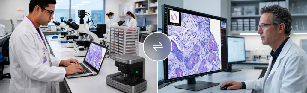
Primaa and PathPresenter Introduce Fully-Integrated AI Workflows for Dermatopathology

Clinical study demonstrates faster, more consistent diagnostic reporting when AI is integrated directly into the pathology viewer; full results presented during September 22 webinar
NEW JERSEY and PARIS, France, September 8, 2026 — PathPresenter, developer of an industry-leading vendor-neutral digital pathology Image Management System (IMS), and Primaa, developer of AI solutions for dermatopathology including the Cleo Skin product, today announced the successful completion of a collaborative validation study on the integration of AI into pathologists’ daily workflow. The results demonstrate improved diagnostic performance as well as faster and more consistent reporting.

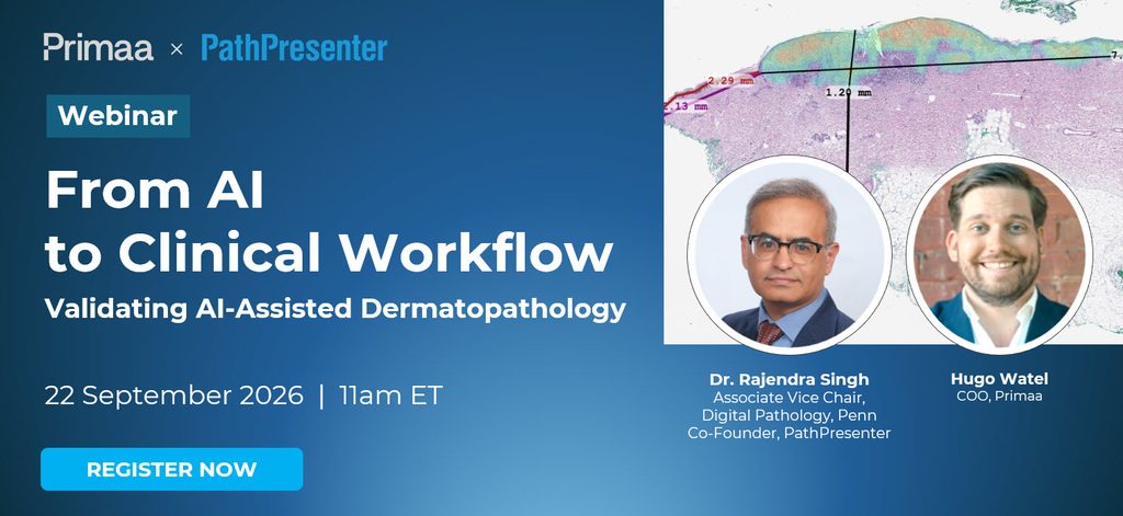
The companies will present the complete study results and demonstrate the validated pathology workflow during a live webinar on Tuesday, September 22, 2026, at 11:00 a.m. Eastern Time.

Unlike many studies that evaluate AI algorithms independently, this research examined AI as part of an integrated diagnostic workflow in which the Cleo Skin analysis appears directly within the PathPresenter viewer, automatically populating structured reporting fields, and enabling pathology reports to be generated with a few keystrokes or even a single click using macro codes. Measurements such as Breslow thickness and mitotic counts are transferred automatically into the report, reducing manual documentation while helping standardize reporting across pathologists. This makes it one of the first AI tools for skin pathology to be tested fully integrated within a digital pathology viewer, with semi-automated reporting built directly into the pathologist’s existing workflow.

Using a two-phase crossover study involving 75 whole-slide images interpreted by seven investigator pathologists, the study compared conventional digital pathology review with the integrated AI-assisted workflow after a four-week washout period.

Cleo Skin is already CE-IVDR certified based on Primaa’s clinical validation studies; this U.S. study complements that clinical evidence, confirming similar results among U.S. pathologists. PathPresenter’s clinical viewer carries both FDA 510(k) clearance and CE-IVDR certification. 

The final results demonstrated measurable improvements in both diagnostic performance and workflow efficiency:

  • 20% reduction in analysis time
  • 17% improvement in diagnostic sensitivity among junior pathologists, highlighting the workflow’s greatest benefit among less experienced readers.

Additional findings, including gains in overall diagnostic sensitivity and immunohistochemistry request rates, will be presented in full during the September 22 webinar.

The study also showed improved performance in melanoma assessment, including reduced Breslow misclassification rates, higher clinical sensitivity, and improved overall diagnostic accuracy.

“Artificial intelligence delivers its greatest value when it becomes a natural extension of the pathologist’s workflow rather than another application to consult,” said Dr. Rajendra Singh, Associate Vice Chair of Pathology at the University of Pennsylvania and PathPresenter co-founder. “This study demonstrates that integrating AI directly into the enterprise image management system can improve both efficiency and diagnostic performance while preserving the pathologist’s central role in clinical decision-making.”

Hugo Watel , COO of Primaa, added, “This partnership reflects why we built Primaa: pathologists are under growing pressure, and AI needs to fit into their workflow to actually help. Integrating Cleo Skin directly into PathPresenter’s platform means it becomes part of pathologists’ daily routine. That everyday use is what makes the impact real: time saved, greater confidence in their practice, and a smoother-running organization.”

Webinar: Final Clinical Results and Workflow Demonstration

During the webinar, Dr. Singh and Mr. Watel will present the study methodology, review the final clinical outcomes, and walk through the integrated workflow used throughout the study. Attendees will gain insight into how AI-assisted reporting can be incorporated into routine dermatopathology practice while improving efficiency and maintaining clinician oversight.

From AI to Clinical Workflow: Validating AI-Assisted Dermatopathology with Primaa and PathPresenter 

Date: Tuesday, September 22, 2026

Time: 11:00 a.m. Eastern Time

Register: https://info.pathpresenter.com/webinar-workflow-primaa 


Live Demonstrations at Pathology Visions 2026

Those interested in seeing these solutions in person are invited to meet with Primaa and PathPresenter for live demonstrations at Pathology Visions 2026 in San Diego, October 16-18, or contact us directly.

About PathPresenter

PathPresenter is the industry’s leading vendor-neutral Image Management System (IMS) for digital pathology. The platform brings together primary diagnosis, remote consultation, image biorepository management, enterprise education, and AI integration in a single interoperable environment, helping pathology organizations streamline workflows while maximizing flexibility, interoperability, and return on investment.

About Primaa

Primaa is a leading provider of integrated AI solutions dedicated to enhance the accuracy and efficiency of biomarker detection in dermatopathology and breast pathology. Its flagship tools, Cleo Breast and Cleo Skin, support pathologists in delivering faster and more reliable diagnoses, ultimately redefining how tissue lesions are assessed.

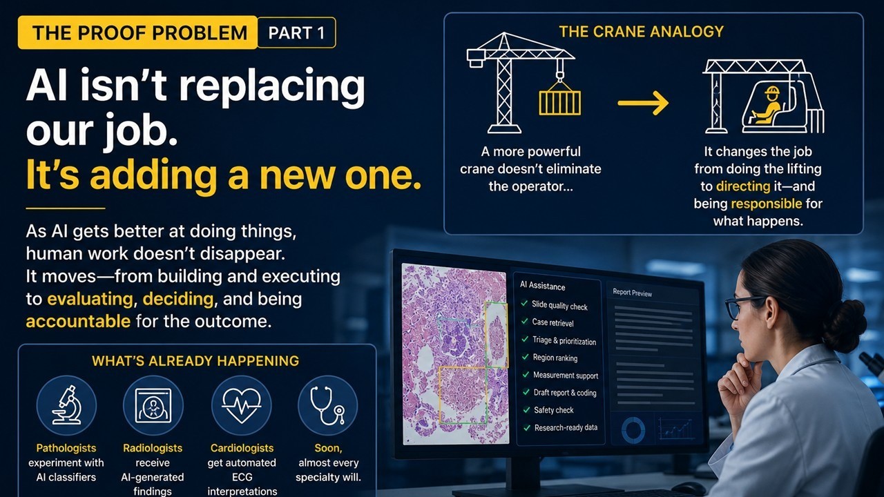
The Proof Problem, Part 1: AI Isn’t Replacing Our Job. It’s Adding a New One.

Every few months, someone tells me AI is coming for pathology. More recently, the message has become reassuring: AI isn’t coming for our jobs, it’s just coming for the boring parts. I don’t think either framing gets the real change quite right.

A recent talk by Princeton computer scientist Arvind Narayanan gave me a different way to think about it. His argument, in short, is that as AI gets better at doing things, human work doesn’t disappear. It moves from building and executing to evaluating, deciding, and being accountable for the outcome. I think that distinction matters enormously in medicine. Narayanan uses the analogy of a crane operator: a more powerful crane doesn’t eliminate the operator; it changes the job from doing the lifting to directing it and being responsible for what happens. In some ways, that may be what is beginning to happen to us.

Think about what is already happening. A pathologist can experiment with a classifier far more easily than a few years ago. A radiologist can receive AI-generated findings. A cardiologist can get an automated ECG interpretation. Soon, almost every specialty will have models sitting somewhere between clinical data and clinical decisions. Building the model is getting easier. Proving that it deserves to influence patient care is not. That may turn out to be one of the most important responsibilities physicians have in the AI era.

Someone still has to ask: Does this model work on our patients? Does it fail differently in different populations? What happens when the scanner, stain, laboratory, protocol, or patient population changes? Is it learning the biology we think it is learning, or exploiting a shortcut we haven’t noticed? And perhaps most importantly, how much should I trust it when there is a real patient on the other side of the prediction? AI can help us do parts of that evaluation, but the standard for what counts as enough evidence, and the accountability for acting on it cannot simply be delegated back to the system being evaluated. That requires clinical judgment.

For years, much of the conversation has been about whether physicians need to learn to build AI. I increasingly think that is only half the question. We also need physicians who know how to validate AI and to determine when, where, and how much it can be trusted. That doesn’t mean physicians shouldn’t learn to build models, or that model development belongs only to engineers and vendors. Quite the opposite: understanding how these systems are built will make us better users and better evaluators of them.

But building and validating are different responsibilities. Physicians have always had to evaluate evidence before acting on it. AI doesn’t create that responsibility from scratch; it expands it. Determining whether an AI system is safe, generalizable, clinically meaningful, and trustworthy is not only a technical question. It is a clinical one too.

And right now, there is a problem: in most health systems, we have not built the infrastructure to do this well. The vendor has evidence. The paper has an AUC. The regulator may have cleared the product. IT can deploy it. But who determines whether it works on our patients, in our workflow, with our instruments, and under the conditions in which we will actually use it?

That is the proof problem, and I suspect solving it will become one of the defining responsibilities of medicine in the AI era.

Over the next few posts, I want to get practical about what that actually means: what to ask an AI vendor, what makes a validation convincing versus what just sounds convincing, how models quietly fail after they’re deployed, and what infrastructure health systems will need before AI can be trusted as part of routine clinical care. I’ll use pathology as my laboratory, because that’s where I sit. But this problem belongs to every specialty, and solving it will require clinicians, scientists, engineers, health systems, regulators, and industry working together.

Next: why “it worked in the paper” might be the least useful sentence in clinical AI, and what we should be asking instead.

This post was prompted in part by Arvind Narayanan’s talk and essay, “What Will Be Left For Us to Work On?” His framing of how AI shifts human work from execution toward evaluation and accountability is well worth reading..

About the Author

Dr. Rajendra Singh is a Professor of Pathology at the University of Pennsylvania and co-founder of PathPresenter. He serves as a member of the Digital and Computational Pathology Committee of the CAP, Editorial Board of the WHO for Classification of tumors, 5th Edition and the Board of Digital Pathology Association.

The Business of Pathology: Unlocking a Lab’s Value

INDUSTRY INSIGHT from Signify Research

Guest Authors:
Alan Stoddart, Editor, Healthcare Technology
Imogen Fitt, Principal Analyst, Digital Laboratory and Lifesciences

Remote consultation solutions offer labs a way to affordably and profitably take advantage of digital pathology

Digital pathology is a discipline of promise.

Providers are promised that the adoption of digital pathology will make them more efficient, enable their pathologists to add greater value and take advantage of sophisticated solutions. These opportunities are real and within reach, but so far only a limited number of sites have been able to realise them.

In many cases, the barriers have been difficult to overcome. The cost of transitioning to digital pathology can be prohibitive, particularly if the return on that investment is hard to quantify. In addition, supporting pathologists, whose priority is their patients, through the change can be challenging for providers. The move can also give headaches to department heads and financial stakeholders, who must cost-effectively navigate issues of interoperability, deploying tools that facilitate flexibility long-term, not simply a solution that works today.

One answer is remote consultation solutions, which offer labs a way to affordably and profitably take advantage of digital pathology, while lightweight implementations minimise adoption challenges.

The Growing Need for Specialty Expertise

Such answers are very timely.

The cases pathology labs are being tasked with are more complex than ever, with growing use of precision medicine leading to more sophisticated testing, while multidisciplinary approaches are driving up the intricacy of a pathologist’s work.

This complexity often benefits from highly specialised expertise, but such expertise is a scarce resource and unequally distributed. However, by using remote consultation solutions, granting labs instant access to a wealth of specialist pathologists from across the world, the impacts of this shortage can be mitigated.

This could be crucial. Without swift, accurate analysis, diagnosis is delayed, which in turn delays treatment planning and delivery.  Delayed or incorrect diagnoses stemming from a lack of expertise not only limit patients’ access to the best possible outcomes, they can be expensive for hospitals. Providers may need to deliver interventions which might otherwise have been avoided or provide costly additional treatments. More starkly, providers are also open to expensive litigation, highlighting the financial risk that a shortage of expertise raises.

Consultation Solutions: The Network Effect

The complexity of conditions, along with the vast range of possible treatments constantly being introduced, means that subspecialty expertise is often required if patients are to benefit from the most personalised care; however, this level of specialisation is often beyond the scope of a single provider.

Remote consultation solutions, such as those enabled by PathPresenter, solve this problem. The company’s tools facilitate secure telepathology for both primary and secondary reads. As such, providers can leverage pathologist capacity outside their own networks, ameliorating constraints and opening access to a large number of specialists regardless of geography.

Labs already using the PathPresenter IMS have remote consultation capability directly available and can leverage PathPresenter’s integration with their existing LIS for workflow.

For those labs yet to fully adopt digital pathology, or for those on IMS systems that lack remote external consultation functionality, there is a zero-footprint version. This fully web-based alternative can be deployed in days without significant investment, LIS integration, or demanding infrastructure requirements. Pathologists can access the solution directly in their browsers via secure web portals, enabling them to receive, view and sign off second opinion cases from referring hospitals across the world.

This lightweight version, in particular, offers laboratories a low-friction route to take their first steps in a digital transformation. With minimal investment, labs can begin to prove the use case of digital pathology on their own patients and build evidence that demonstrates a return on investment, allowing providers to make better-informed decisions as they ramp up their rollout of digital pathology.

In doing so, providers can leverage the advantages that digital consultation workflows support. Pathologists are better able to devote themselves to the parts of their work which add the most value, with resource limitations and the pressures they create mitigated. They are also better able to leverage their specialist expertise, providing valuable second opinions on complex cases while also generating revenues for their own pathology departments. As such, remote consultations and second opinions have the potential to become a key tool in every institution’s arsenal.

The Future Pathology Department and the Pathology Feedback Loop

The availability of this capability will help transform pathology departments in the coming years.

Patients and their physicians will not be limited by the knowledge within their own institutions. Instead, advanced subspecialty expertise can be enlisted, augmenting a provider’s own in-house team to deliver more personalised care and improved outcomes. Referring institutions will easily and affordably be able to leverage this expertise, thanks to low-impact tools that make recruiting external pathologists simple.

Effectively, this also increases the utility of subspecialist expertise. By making it more accessible, its value is increased, thereby justifying greater resources devoted to this expertise. This positive feedback loop empowers pathologists’ own development, allowing them to specialise further; it is a valuable asset for providers as they meet the demands of precision medicine.

Perhaps the most transformative result will come at an enterprise level, however. Pathology is, at present, often considered a necessary expense. Its value to patient care is unquestioned, but it is a cost centre, not a profit centre, impacting the department’s influence and investment. Remote consultation can be instrumental in overcoming this, and providing second opinions for other institutions can generate hundreds of dollars a case. Given the relatively low investment required to adopt PathPresenter’s solutions and realise these revenues, offering second opinions could be a quantifiable income stream for institutions, as organisations such as The Ohio State University Comprehensive Cancer Center-Arthur G. James Cancer Hospital and Cincinnati Children’s Hospital Medical Center have found.

This value justifies increased investment in pathology departments, enabling procurement of additional hardware and software solutions, as well as investment in new technologies such as AI. In doing so, remote consultation can become a core component in the elevation of in-institution diagnostics. 

Pathologists themselves also stand to benefit, with their roles evolving as a result of consult tools. For instance, PathPresenter’s browser-based platform makes it easier for pathologists to work flexibly and remotely. Instead of relying almost entirely on the slide itself, remote consult solutions also mean that diagnosticians will have much more information available to them, and advanced AI diagnostic tools can be applied more directly. This will position pathologists more centrally in multimodality diagnostic workflows, ultimately empowering them to take a lead role in the realisation of precision medicine.

Delivering Pathology ROI

Realising this vision of a profitable, efficient, pathologist-centred laboratory is in reach, but adopting these processes will require active management on the part of institutions. Expensive, admin-heavy and time-consuming consultation practices involving the preparation, protection and physical sharing of slides can be replaced by digital delivery. This makes the process cheaper, near instantaneous and with reduced risk of damaging the slide. It also enables the same sample to be assessed by several pathologists simultaneously, should varied expertise be required.

Remote consultation solutions’ role in addressing these barriers, lowering these costs and mitigating these risks also aids the transformation of pathology departments from cost centres into profit centres, which further enables their evolution. Vendors such as PathPresenter could be especially valuable in this process. The vendor’s IMS helps pathology departments already deep in their digital pathology transformation to capitalise. But those who have not yet made such a deep investment can still take advantage of the vendor’s lightweight platform. This may be a gentler route to unlocking the opportunities offered by digital pathology, enabling practical success and building value before financial commitment is needed.

Above all, the greatest promise offered by digital pathology’s adoption is in granting patients longer, healthier lives. Transitioning to the technology has usually represented an expensive shift for providers, with significant risks. But, a lightweight, flexible approach flattens these hurdles, enabling digital pathology to be utilised, without a daunting commitment.

Moreover, remote consultation solutions such as those offered by PathPresenter mitigate shortages of pathologists and allow the expertise of subspecialist pathologists to be leveraged as a central component of multidisciplinary workflows. In doing so, they help providers to truly realise digital pathology’s promise. 

— — —

About the Authors

Alan Stoddart joined Signify Research in 2020. Coming from a business intelligence background, Alan now leads Signify Research’s Insight and Editorial services. His work focuses on Signify Research’s Digital Laboratory & Life Sciences Premium Insight service, where he covers digital pathology, laboratory technologies, and healthcare innovation through independent market analysis and industry insights.

Imogen Fitt is a Principal Analyst at Signify Research, leading the company’s coverage of Digital Laboratory and Lifesciences markets. Since joining in 2018, she has been instrumental in establishing Signify Research as a leading authority in digital pathology market intelligence. She holds a First-Class degree in Biomedical Sciences from the University of Warwick and brings additional expertise across Laboratory Information Systems, genomics, real-world data, and AI in drug development, supporting expanded coverage of both clinical and preclinical markets.

Expanding Access to Hantavirus Education: Sharing Images Through the PathPresenter Public Library

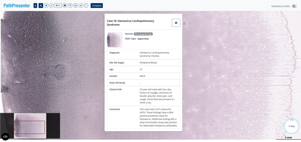
What does hantavirus look like to a pathologist? On a digital whole slide image of a peripheral blood smear, it looks a lot like this.

Hantavirus is a rare but serious infection, and the recent headline-grabbing surge in cases around the world highlights the importance of timely diagnosis in preventing its spread. Recognition of hantavirus may begin with clinical teams seeing symptoms, but pathologists and laboratory professionals play a key role in identifying subtle morphologic features in peripheral blood smears, leading to confirmatory testing and patient management. With this in mind PathPresenter is proud to highlight the work of Dr. Joseph Stenberg, a resident at the University of New Mexico School of Medicine, who is sharing an educational series focused on hantavirus cardiopulmonary syndrome (HCPS) screening in peripheral blood smear cases.

The project comprises 10 hand-selected cases by Dr. Kathryn Foucar, Distinguished Professor at the University of New Mexico, and includes whole slide images (WSIs) representing a broad spectrum of diagnostic presentations. These include classic examples of HCPS, borderline cases, early-evolving infections, and negative mimics that challenge learners to distinguish subtle findings from true disease.

By sharing cases across this diagnostic spectrum, Dr. Stenberg aims to create a valuable educational resource for pathology trainees, practicing pathologists, and other laboratory medicine professionals seeking to improve their recognition of hantavirus-related findings in peripheral blood smears.

Bringing Interactive Learning to Published Medical Education

A key goal of the project is to allow learners to do more than simply view static images. To maximize educational value, Dr. Stenberg is using PathPresenter to provide an interactive slide-viewing experience.

“This project was built around a simple goal: to make high-quality educational cases of hantavirus cardiopulmonary syndrome freely available to anyone, anywhere” said Dr. Stenberg. “By providing unrestricted access to complete digital slides through PathPresenter, we hope to help laboratory professionals become more confident in recognizing this rare but life-threatening syndrome. Earlier recognition can lead to earlier diagnosis, more timely clinical intervention, and ultimately better patient care.”

Rather than relying on selected screenshots or figure panels alone, learners can explore the complete digital slides themselves, zooming and navigating through the specimens just as they would at a microscope. This approach allows users to appreciate subtle findings, evaluate areas of interest independently, and gain experience that more closely resembles real-world diagnostic practice.

To make the cases as accessible as possible, Dr. Stenberg has generously donated de-identified slides to the PathPresenter Public Library. The slides will also be linked from his forthcoming publications (both online and in print), allowing readers to move seamlessly between published educational content and the original digital pathology slides.

A Great Example of Community Contribution

Dr. Stenberg’s project exemplifies one of the core purposes of the PathPresenter public platform: creating a collaborative learning community where educators and learners can contribute to the advancement of pathology education.

Any PathPresenter public user can share de-identified educational material with the community. Contributions from individual pathologists, trainees, academic institutions, and professional organizations collectively help build a richer and more diverse educational ecosystem.

Many users are familiar with PathPresenter’s highly popular High Yield Cases, which are curated and annotated by distinguished pathologists and serve as valuable study resources for residents preparing for board examinations. But the continued growth and success of the platform is driven by contributions from across the pathology community.

Today, the PathPresenter public platform includes more than 85,000 users and over 10,000 slides, representing a broad range of subspecialties, diseases, and educational approaches. Every new contribution helps expand the breadth of learning opportunities available to pathologists around the world.

A Valuable Resource for Hematopathology Education

The HCPS cases contributed by Dr. Stenberg consist of blood smear (hematopathology) images scanned at 40× magnification, providing the resolution needed to examine important cellular details and morphologic features.

Because the collection includes both classic and challenging presentations, it offers learners an opportunity not only to recognize hallmark findings but also to develop diagnostic judgment in cases where the answer may not be immediately apparent. These are often the cases that provide the greatest educational value and most closely reflect real-world practice.

As his cases are published, they will serve as a useful bridge between traditional academic publication and interactive digital pathology education.

Thank You, Dr. Stenberg

PathPresenter salutes Dr. Stenberg and his colleagues for this valuable contribution to publicly available medical knowledge. By sharing these cases with the pathology community, they are helping create resources that will benefit learners and educators far beyond their own institution.

We look forward to sharing links to Dr. Stenberg’s published articles in the near future and will post updates here on the PathPresenter blog as they become available.

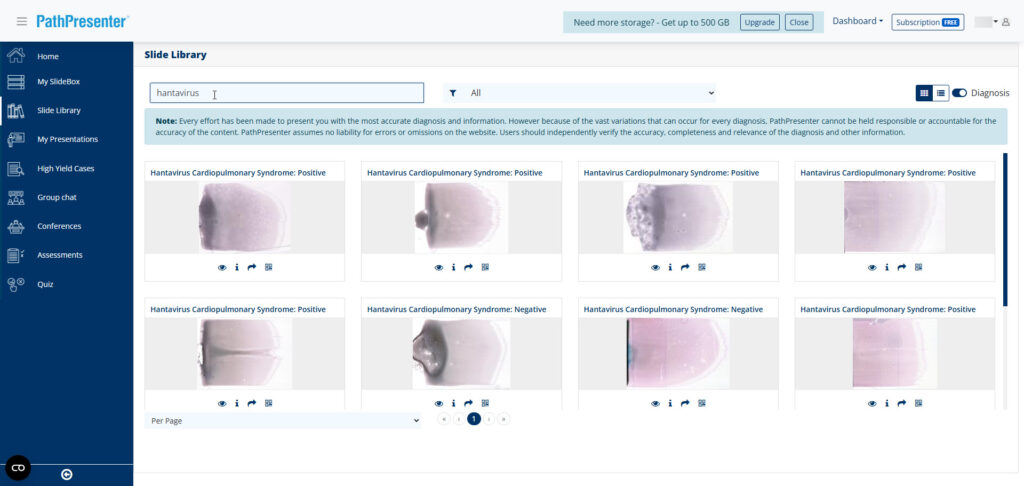
In the meantime, if you’d like to explore the slides yourself, visit the PathPresenter Public Library and search for “hantavirus.” If you don’t already have one, you can create a free account which includes 10 GB of storage for your own uploads. 

Hantavirus slides in the PathPresenter Public Library

We encourage educators, trainees, and practicing pathologists alike to take advantage of these interactive teaching materials—and to consider contributing their own educational cases to help strengthen the global learning community.

The Future of Pathology Part 3: The Delivery Problem and the Infrastructure of Intelligence
After Part 1 and Part 2, many of you asked how the argument ends. This is where it lands.

When a field reaches consensus, it usually means the real work is about to begin.

Over the past two essays, I argued that the greatest challenge in precision oncology is no longer discovery. We continue to identify powerful biomarkers, develop increasingly sophisticated AI models, and generate molecular insights at an unprecedented pace. Scientific discovery creates possibility. Delivery creates outcomes.

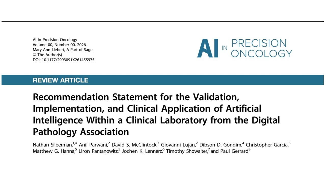
This week, the Digital Pathology Association released a comprehensive recommendation statement on the validation, implementation, and clinical application of artificial intelligence in digital pathology. What struck me was not what was new, but what it quietly confirmed. The document is nominally about artificial intelligence. What it really describes is the operating environment required for precision medicine.

Three recommendations reveal just how much the conversation has changed.

1. Variability

The first concerns scanner variability. The article is explicit: algorithm performance depends on the scanner on which it is deployed, and validation must be demonstrated for each platform. This is more than a technical consideration and an acknowledgment of operational reality. Hospitals rarely operate identical hardware, workflows, or laboratory environments, and precision medicine must succeed despite that variability, not because it has been eliminated.

2. Oversight

The second concerns pathologist oversight. The recommendation is unequivocal: AI augments clinical expertise but does not replace it. In daily practice, this feels less like a regulatory requirement than a recognition of clinical reality. While AI excels at narrowly defined tasks, pathologists must integrate morphology, clinical history, molecular findings, rare disease patterns, and diagnostic judgment that no training set fully anticipates. The future is not autonomous diagnosis. It is intelligently augmented expertise.

3. Clinical Utility

The third concerns clinical utility, and this is where the bar is raised most significantly. A high-performing model is not enough. Clinical utility requires evidence that using the result actually changes patient management and improves outcomes. In dermatopathology, where a melanoma grading decision can determine treatment intensity, that distinction is not abstract. It moves the conversation beyond developing algorithms to delivering measurable clinical value, and that is a fundamentally harder problem.

Taken together, these recommendations describe something much larger than AI governance. They describe the infrastructure required for intelligence to become healthcare.

We have seen this pattern before. GPS technology existed for decades, yet it did not transform everyday life simply because satellites became more accurate. It became indispensable only when maps, smartphones, cloud computing, wireless networks, and real-time traffic data converged into a seamless ecosystem. The breakthrough was not a better satellite. It was the infrastructure that allowed millions of people to benefit from the intelligence those satellites had been generating all along. Precision oncology is approaching a similar inflection point. The biomarkers exist, the algorithms exist, and our biological understanding continues to expand. What remains inconsistent is the operational layer that validates performance across diverse environments, orchestrates workflows, connects fragmented systems, governs quality, and delivers the right intelligence at the precise moment a clinical decision must be made.

The conversation is evolving. We are moving from asking “Can AI detect disease?” to asking “What system ensures that AI improves care, every case, every day?” That is a much more difficult question, and it is the one that will determine whether precision medicine scales beyond a handful of highly resourced institutions or becomes accessible to patients everywhere.

Discovery will continue to accelerate. But the breakthroughs that matter are the ones that become routine, and that requires infrastructure, execution, and a field willing to treat delivery as seriously as discovery.

About the Author

Dr. Rajendra Singh is a Professor of Pathology at the University of Pennsylvania and co-founder of PathPresenter. He serves as a member of the Digital and Computational Pathology Committee of the CAP, Editorial Board of the WHO for Classification of tumors, 5th Edition and the Board of Digital Pathology Association.

What ASCO Taught Me About the Future of Pathology – Part 2

TROP2, Tissue, and the Missing Spatial Dimension

Last week, I wrote that the future of pathology would not be defined by any single technology—not AI, not molecular testing, and not digital pathology alone. Rather, the future belongs to those who can read tissue morphology, imaging, molecular biology, and clinical outcomes as a single integrated story rather than four separate chapters.

The discussions that followed were illuminating. Conversations with radiologists, oncologists, data scientists, and colleagues across the pharmaceutical industry reinforced something I had already begun to suspect: many of the most important questions in precision medicine no longer sit comfortably within the traditional boundaries of a single specialty.

As I reflected on the clinical data presented at ASCO this year, one story stood out because it illustrates exactly where this convergence may be heading. That story is TROP2.

Why TROP2 Matters: The Architecture of an ADC

For those not immersed in oncology drug development, TROP2 (trophoblast cell-surface antigen 2) is a transmembrane glycoprotein expressed across a wide range of epithelial malignancies, including lung, breast, bladder, and endometrial carcinomas. It has become a major target in oncology because it serves as an address for a rapidly expanding class of therapies known as antibody-drug conjugates (ADCs).

The concept is elegant. An antibody binds a target on the cancer cell, carries a highly potent cytotoxic payload, and delivers that payload into the cell following binding and internalization. In practice, many ADCs—including TROP2-directed therapies—also exert a “bystander effect,” where payload can diffuse into neighboring cells, making therapeutic activity less dependent on perfectly uniform target expression.

At ASCO, clinical data across multiple programs continued to reinforce the potential of this strategy. Early-phase and emerging late-phase results suggest meaningful clinical activity for TROP2-directed ADCs, including in combination with immunotherapy in selected settings. With multiple global trials underway across tumor types, this represents one of the most active areas of drug development in oncology today.

There is clear biology here—and real benefit for patients who respond. Yet as these data accumulate, a fundamental question becomes harder to ignore: how do we determine which patients will benefit most? The answer may expose a broader limitation in how we currently define biomarkers.

What the H-Score Misses

Today, our approach to TROP2 remains centered on expression. Pathologists stain tissue, evaluate intensity and extent, and report a score. Computational approaches have improved on this by quantifying expression more reproducibly and, in some cases, distinguishing subcellular localization.

These are meaningful advances. But they are still asking the same core question: What is the biology of TROP2?

At the microscope, the question often feels different: What is the biology surrounding TROP2?

A biomarker is never seen in isolation. Tumor cells exist within a structured environment: immune cells attempting to infiltrate, fibroblasts shaping stromal architecture, and physical barriers organizing the tumor into compartments. The tumor–stroma interface—the boundary between malignant epithelium and host response—is often where critical biology resides.

Experienced pathologists recognize these patterns intuitively. When I am teaching residents, I remind them that the tissue is often smarter than the pathologist. If the stroma is walling something off, that reaction is telling you something. The interface between tumor and host is rarely silent. Yet much of this contextual information is lost when complex tissue architecture is reduced to a single expression score.

That raises an important possibility: What if response to TROP2-directed therapy depends not only on target expression, but also on the biological context in which that target exists?

The Missing Spatial Dimension: An Ecosystem Approach

Consider the immune microenvironment.

A tumor with high TROP2 expression but minimal immune infiltration may behave very differently from one with the same expression profile embedded within an organized immune response. The molecular target may be identical, but the surrounding ecosystem is not.

Preclinical and early clinical observations suggest that ADC activity may interact with the tumor microenvironment in ways that extend beyond direct cytotoxicity. If those interactions contribute to clinical response, then baseline tissue architecture—including immune organization—becomes an important variable that current biomarker strategies only partially capture.

This concept is not new. In colorectal cancer, the Immunoscore demonstrated that the spatial distribution of immune cells—particularly at the invasive margin—has strong prognostic value and has influenced how we think about tumor–immune interactions more broadly.

Yet we have only begun to ask whether similar spatial features influence response to ADCs such as those targeting TROP2.

Even at a purely physical level, tissue architecture may matter. A TROP2-high tumor with dense stromal compartmentalization could experience very different drug delivery and payload diffusion dynamics than a tumor with a more permissive structure. These are measurable features—but they are rarely incorporated into current biomarker frameworks.

This points toward a broader realization: we may have spent years searching for answers in individual molecules when the more informative signal, in some settings, lies in the relationships between them—their spatial arrangement, biological context, and interaction within a tissue ecosystem.

Beyond the Slide

The second observation that stayed with me after ASCO is that biology does not operate at a single scale.

Tumors do not live on glass slides. They exist within tissues, tissues within organs, and organs within patients. It follows that response to therapy is shaped across all of these levels simultaneously.

Some relevant features are visible histologically. Others emerge through imaging, molecular profiling, or longitudinal clinical data. Increasingly, computational approaches are making it possible to integrate these layers.

The central question for precision oncology is no longer which modality is most informative in isolation. It is whether these modalities are capturing different dimensions of the same biological system—and whether we are building the infrastructure to interpret them together.

For decades, medicine has organized these data into separate workflows, databases, and specialties. Biology never made those distinctions.

The Bigger Lesson

TROP2 is not the story. It is a case study.

For the past two decades, precision medicine has focused on identifying the right molecule—and that approach has transformed cancer care. But in some contexts, the molecule alone may not be sufficient to explain therapeutic response.

Tumors exist within tissues. Tissues exist within organs. Organs exist within patients. Every level contains information. Every level influences outcome.

This is why pathology is becoming more important—not less—in the era of AI and advanced molecular diagnostics. Not because it replaces other disciplines, but because it is inherently grounded in biological context.

The role of the pathologist is evolving accordingly. It is no longer limited to interpreting a slide or assigning an expression score. It increasingly involves connecting tissue architecture, molecular data, imaging, and clinical outcomes into a more complete model of disease.

That integration is beginning to take shape. The groups that build this connective infrastructure first will be positioned to answer some of the most important biomarker questions in oncology.

The tissue has been telling us this story for years. Imaging has been telling the same story from a different vantage point. Molecular data and clinical outcomes add additional layers. For most of modern medicine, we have read these stories separately.

We are finally learning how to read them together.

About the Author

Dr. Rajendra Singh is a Professor of Pathology at the University of Pennsylvania and co-founder of PathPresenter. He serves as a member of the Digital and Computational Pathology Committee of the CAP, Editorial Board of the WHO for Classification of tumors, 5th Edition and the Board of Digital Pathology Association.

What ASCO Taught Me About the Future of Pathology

A field report from the world’s largest oncology meeting—and a call to my fellow pathologists.

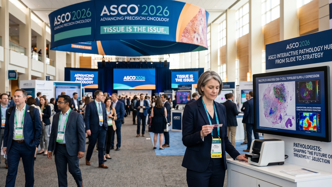
This weekend, I joined tens of thousands of oncologists, researchers, pharmaceutical executives, and data scientists at the ASCO Annual Meeting. The entire cancer medicine ecosystem was gathered in one place.

Yet among the thousands of people discussing the future of cancer care, I could count the pathologists on two hands.

I sat through presentations on antibody-drug conjugates (ADCs), immunotherapy combinations, spatial biomarkers, and novel companion diagnostics. Different diseases. Different companies. Different therapeutic strategies. Yet I found myself writing the same note repeatedly in the margin of my notebook:

This depends on pathology.

The more I listened, the more obvious it became that many of oncology’s biggest challenges are no longer drug problems. They are patient-selection problems. And patient selection begins with understanding tissue.

The industry is chasing the biology that pathologists have spent their careers studying.

For the first time in my career, I believe pathology is positioned not only to diagnose disease, but to help determine treatment.

1. The Industry Is Chasing Tissue Biology

No matter the disease or the drug class, the central question in oncology today is patient selection. Who will respond? Who will benefit? Who should receive this therapy?

Whether the discussion involved checkpoint inhibitors, ADCs, or cellular therapies, the answers do not live in a liquid biopsy or a genomic sequence alone. They live in tissue architecture inside the tumor microenvironment, within the spatial relationships between malignant cells and immune infiltrates.

The answers oncologists are looking for reside in tissue. And that means pathology must be at the table.

2. The H&E Slide Is Far Richer Than We Imagined

The glass slide sitting under our microscopes contains a vast reservoir of unextracted information. Spatial arrangements of immune cell subsets, subtle variations in stromal organization, cellular heterogeneity across a specimen are not incidental features. They are biological signals that influence treatment response and clinical outcomes.

Digital pathology, computational modeling, multiplex imaging, and AI allow us to quantify what was previously qualitative, transforming what we have always observed into measurable, reproducible biological data.

These technologies are not replacing the pathologist’s eye. They are extending its reach. This is not a disruption to pathology; it is pathology at greater resolution.

3. Pathology’s Second Act: From Diagnosis to Treatment

For most of my career, pathology has answered the first question in cancer care: What disease does this patient have? Oncology is now asking a second question: What treatment is most likely to help this patient?

That question increasingly depends on information embedded within the tissue itself:

  • Why does one patient respond to immunotherapy while another progresses?
  • Which features of the tumor microenvironment predict resistance?
  • What makes a targeted therapy effective in one patient and ineffective in another?

These are pathology questions. And they are among the most consequential questions in medicine today.

4. Why Pathologists Must Lead

No algorithm inherently understands tissue biology. No software comprehends the clinical weight of a diagnostic error. Pathologists do. That is precisely why we must be the ones evaluating, validating, and challenging these tools, separating what is biologically meaningful from what is merely statistically interesting.

The pathologist of the next decade will need an expanded toolkit. We must learn how to interpret spatial biology, understand AI-derived biomarkers, engage with computational pathology, and participate in the development of the next generation of companion diagnostics. This is not about abandoning what we know; it is about building on it.

The future of oncology cannot be built by software engineers or clinical oncologists alone. It requires pathologists who speak both the language of tissue and the language of data.

The future of pathology will not be determined by artificial intelligence. It will be determined by whether pathologists choose to engage with it.

The tools are changing. The mission is not.

Defining the Future

When I return to ASCO next year, I hope I need more than two hands to count the pathologists in attendance. Not because we are defending our relevance, but because we are helping shape the future of cancer care.

The future of precision oncology is being built now.

And pathology belongs at the center of it.

About the Author

Dr. Rajendra Singh is a Professor of Pathology at the University of Pennsylvania and co-founder of PathPresenter. He serves as a member of the Digital and Computational Pathology Committee of the CAP, Editorial Board of the WHO for Classification of tumors, 5th Edition and the Board of Digital Pathology Association.

A Biomarker by Any Other Name Would Smell as Sweet: Building an AI-Powered Internal Concept Library

Part two of a two-part series on the idea of Internal Concept Libraries. Read part one.

Building an AI-powered internal concept library

The promise of artificial intelligence in pathology is often framed around image analysis and diagnostic augmentation. But one of the most immediate opportunities may lie elsewhere: transforming decades of unstructured pathology reports into computable, searchable clinical data.

A recent pathology informatics project by Raj Singh, Professor of Pathology at UPenn and PathPresenter co-founder, and Alexander Goel, COO at PhenoML, explored this challenge through the development of an AI-driven extraction pipeline designed around a central principle: preserve all clinically meaningful observations, even when standardized terminology systems fail to represent them completely.

The project focused on constructing an Internal Concept Library capable of bridging the gap between narrative pathology reporting and structured research databases.

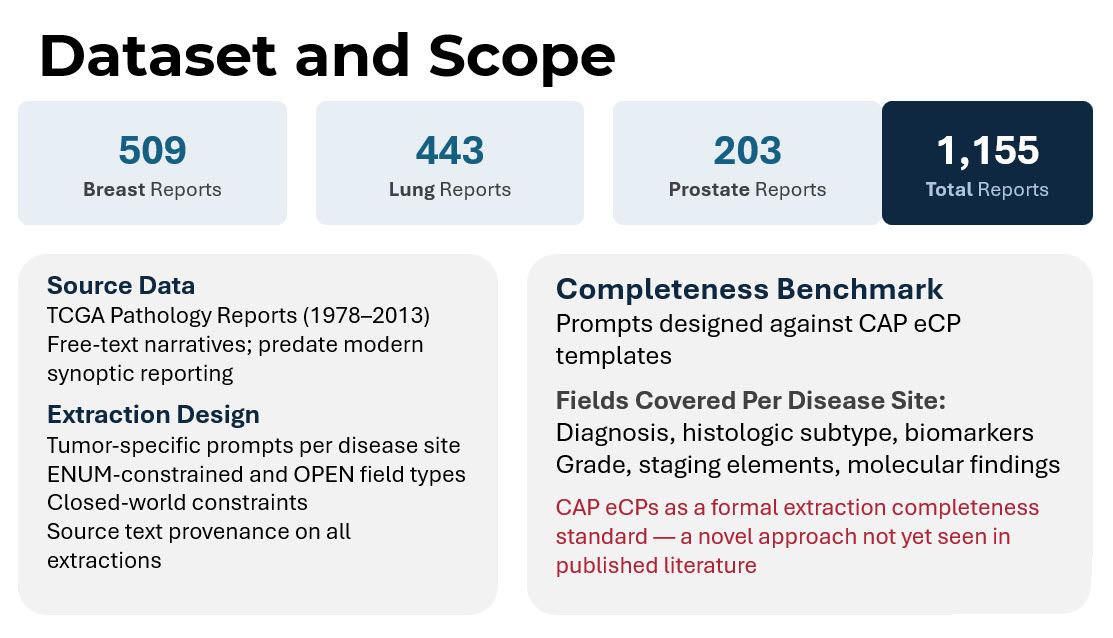
The dataset consisted of 1,155 free-text pathology reports drawn from The Cancer Genome Atlas (TCGA), including breast, lung, and prostate cancer cases spanning 1978 to 2013. These reports largely predated modern synoptic reporting practices and therefore represented the type of highly variable narrative text still common throughout historical pathology archives.

The objective was not simply to extract data fields. It was to evaluate whether a concept-first architecture could preserve more clinically useful information than traditional terminology-dependent pipelines.

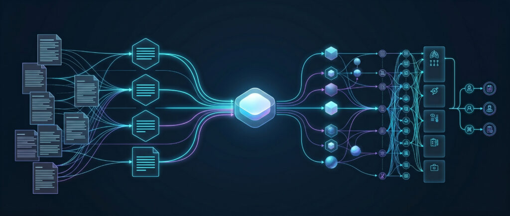
The system architecture was intentionally designed to separate clinical meaning from terminology standardization.

The pipeline began with ingestion of raw pathology reports. Cases were first routed through disease-specific classification using external terminology APIs and machine learning classifiers. Each report was then processed using large language model prompts tailored to individual disease sites.

Rather than allowing unconstrained extraction, the prompts used a closed-world design philosophy. The model was explicitly instructed that the field list was exhaustive, preventing the AI from hallucinating expected findings that were not actually documented in the report.

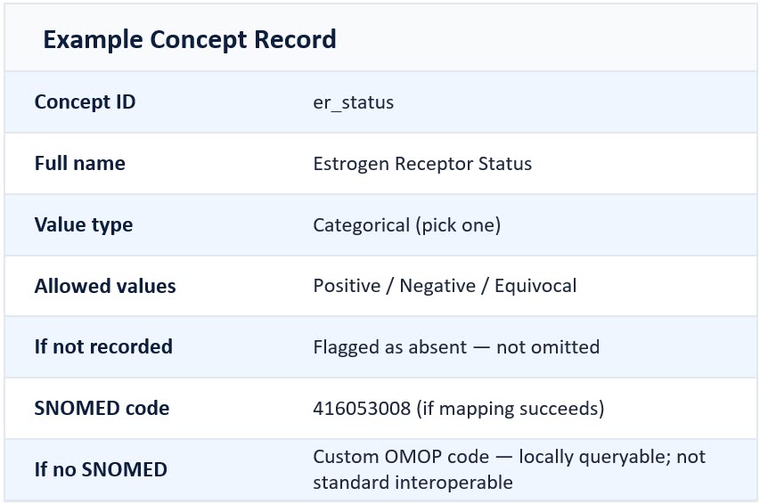
Each extracted observation was assigned an internal concept identifier before any attempt at external terminology mapping occurred. This architectural decision represented the core innovation of the project.

In traditional pipelines, extraction is often tightly coupled to successful SNOMED encoding. If no suitable standard code is found, the observation may be excluded from downstream datasets entirely.

In the concept-library model, however, the internal concept persists regardless of SNOMED availability.

Only after extraction were findings submitted to terminology matching systems to identify corresponding SNOMED CT or OMOP standard concepts. When no standard code existed, custom OMOP concept IDs were assigned instead, preserving full queryability within the OMOP ecosystem. The results highlighted the scale of the terminology gap.

Study results
Results of extraction and mapping

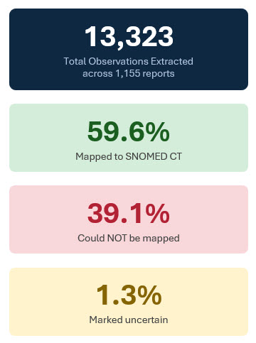
Across 1,155 reports, the system extracted 13,323 clinical observations. Of these, approximately 59.6% successfully mapped to SNOMED CT concepts. Roughly 39.1% could not be mapped to standard terminology despite representing clinically meaningful findings. An additional 1.3% were flagged as uncertain due to ambiguity or conflicting evidence within the report text.

Under conventional code-first architectures, a substantial portion of these observations would likely have been excluded from structured datasets entirely. Instead, the Internal Concept Library preserved all extracted findings.

The project also evaluated extraction completeness against the College of American Pathologists electronic Cancer Protocols (CAP eCPs), using the CAP templates as a formal benchmark for information coverage. This approach itself represented a novel methodological contribution, as CAP templates have rarely been used as explicit completeness standards in published large language model pathology extraction research.

Coverage varied by disease site. Breast reports achieved approximately 32.9% CAP field extraction, prostate reports 36.5%, and lung reports 21.8%, with an overall completeness rate of 29.2%.

At first glance, those percentages may appear modest. But the context matters enormously.

The TCGA reports originated from an era before structured synoptic reporting became widespread. Many CAP-defined fields simply did not exist within the source narratives. In other words, lower completeness often reflected absence of documentation rather than extraction failure.

The researchers hypothesized that applying the same pipeline to contemporary synoptic or semi-structured pathology reports would likely produce substantially higher completeness rates.

Several important design lessons emerged from the project.

  • First, closed-world prompting proved essential. Explicitly constraining the extraction schema reduced over-extraction and prevented the model from inferring clinically plausible but undocumented findings.
  • Second, the team found that self-reported LLM confidence scores were poorly calibrated and unreliable for quality assurance. Instead, better performance indicators emerged from structured assertion logic (e.g. forcing every extracted finding into explicit states such as present, absent, or uncertain rather than allowing vague narrative interpretation), explicit review of terminology concordance (verifying that extracted findings align consistently with accepted clinical vocabularies), and cross-field consistency checks (evaluating whether extracted observations remain clinically coherent when considered together).
  • Third, the researchers intentionally avoided creating separate post-extraction normalization stages. Additional normalization layers often compound error propagation while duplicating functionality already present in terminology APIs.

Most importantly, the study demonstrated that pathology AI systems should prioritize preservation of meaning over immediate standardization.

This distinction has major implications for clinical trial recruitment and translational research. Using concept-level querying, researchers could identify highly specific patient cohorts using combinations of biomarkers, grading systems, and morphologic findings regardless of whether standardized terminology mappings existed for every observation.

Queries such as:

  • Triple-negative breast cancers with Ki-67 greater than 50%
  • Gleason score ≥8 with perineural invasion

could operate across the full extracted dataset, not merely the subset successfully encoded in SNOMED.

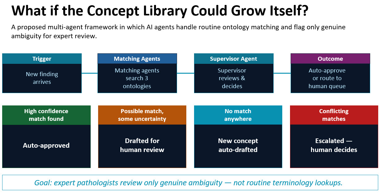
The project also pointed toward future directions in ontology management itself. Singh and Goel are considering multi-agent AI frameworks capable of automatically reviewing new observations, searching multiple ontologies, drafting candidate mappings, and escalating only genuinely ambiguous cases for human expert review. In such systems, pathologists and informaticians would spend less time performing repetitive terminology lookups and more time adjudicating clinically meaningful ambiguity.

Internal Concept Library multi agent framework

Ultimately, the work reinforces a broader shift occurring across healthcare AI. The goal is no longer simply extracting structured data from pathology reports. The larger challenge is preserving clinical meaning at scale while enabling interoperability, research, and machine reasoning.

Pathologists have always documented nuanced clinical interpretation for human readers. Building systems that preserve that same nuance for computational systems may become one of the defining informatics challenges of precision medicine.

What’s in a Namespace: The Critical Role of an Internal Concept Library for Scalable Pathology Data Management

Part one of a two-part series on the idea of Internal Concept Libraries

Internal Concept Library

Modern pathology departments generate enormous volumes of clinically rich information every day. Yet much of that information remains effectively invisible to the systems used for identifying patients for research studies, biomarker-driven therapies, and clinical trials.

Much of the problem stems from a lack of structure.

Across healthcare institutions, pathology reports are still dominated by unstructured narrative text: highly expressive for human interpretation, but difficult for computers to interpret consistently. As precision medicine increasingly depends on identifying patients with highly specific molecular and histologic characteristics, this gap between human-readable and machine-readable pathology is becoming one of the most important bottlenecks in translational research. It prevents modern tools from leveraging critical data at scale.

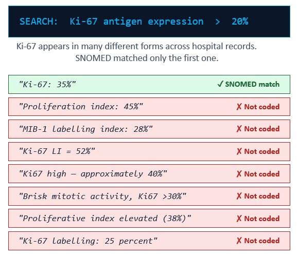
At the 2026 Association for Pathology Informatics Summit, Raj Singh, Professor of Pathology at UPenn and PathPresenter co-founder, asked the audience to consider a clinical trial scenario: A researcher needs to identify patients with triple-negative breast cancer whose Ki-67 proliferation index exceeds 20%. Querying their institution’s database using the standard SNOMED CT concept associated with Ki-67 expression returns 47 patients.

The problem? The actual number of eligible patients in the database is 112.

Sixty-five patients were missed—not because the information was absent, but because it was described differently across pathology reports.

One report may say “Ki-67: 35%.” Another may describe “Proliferation index: 45%.” Others may reference “MIB-1 labeling index,” “Ki67 high,” or “Brisk mitotic activity.” To a pathologist, these phrases convey closely related clinical meaning. To a conventional search system relying on standardized terminology mapping, many are effectively invisible.

Ki-67 search results

This challenge is not unique to Ki-67. It reflects a broader reality in pathology reporting.

Different institutions, and even different pathologists within the same institution, use varying terminology, abbreviations, units of measurement, and narrative styles. A tumor described as “2.2 cm” in one report may appear as “22 mm” in another and “0.022 m” in a third. Humans easily recognize these as equivalent observations. Most database search tools do not.

The industry’s primary solution to this problem has been terminology standardization frameworks such as SNOMED CT and the OMOP Common Data Model (CDM). These systems are essential foundations for interoperability and large-scale clinical research, and they are both impressive and commendable. But they also expose an important limitation: medicine evolves faster than standardized terminology systems can fully capture.

SNOMED CT (Systematized Nomenclature of Medicine Clinical Terms) is one of the world’s most comprehensive clinical vocabularies. It provides unique identifiers for clinical concepts so that systems across institutions can interpret observations consistently. When a pathology finding is successfully mapped to SNOMED, that information becomes interoperable and searchable across research networks.

OMOP CDM, meanwhile, provides a standardized database structure widely used by the OHDSI research community. OMOP enables federated research across hundreds of institutions while preserving patient privacy. Importantly, OMOP also supports custom local concepts through reserved concept ID namespaces. In other words, it explicitly leaves space in its system for concepts that haven’t been defined yet, knowing that medicine and research are always expanding. 

Together, SNOMED and OMOP form the backbone of many modern clinical data infrastructures. But they are not sufficient on their own. 

One core issue is that traditional pathology extraction pipelines often treat successful terminology mapping as a prerequisite for data preservation. If an extracted observation cannot be confidently mapped to a standard code, it is frequently discarded from downstream datasets entirely. Even if the data is preserved in local OMOP custom concepts, those local concepts do not provide any shared meaning across institutions.

This creates a dangerous form of silent data loss. The pathologist documented the finding correctly. AI extraction systems may even identify it correctly. But if no standard code exists, or if terminology matching fails, the information disappears from searchable research datasets.

For institutions engaged in clinical trials, biomarker research, or retrospective cohort studies, this means potentially eligible patients are never identified.

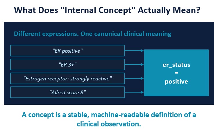
The solution proposed by Dr. Singh, in collaboration with Alexander Goel, COO at PhenoML, is the development of an Internal Concept Library. An internal concept is one stable name for one clinical observation: it gives a consistent way for people and systems to recognize it, regardless of how the pathologist wrote it. An Internal Concept Library acts as a canonical semantic layer between unstructured pathology language and external terminology systems.

For example, multiple report expressions such as:

  • “ER positive”
  • “ER 3+”
  • “Strongly reactive estrogen receptor”
  • “Allred score 8”

can all resolve to a single internal concept:

er_status = positive

The internal concept becomes the persistent primary key. Standard terminology mappings are then attached downstream when available. Instead of treating SNOMED codes as the primary identifiers, this approach first maps observations to stable internal concepts designed specifically around clinical meaning.

This architectural distinction is critically important. If SNOMED mapping succeeds, the observation gains full interoperability. If SNOMED mapping fails, the observation is still preserved, queryable, and available for cohort discovery. Nothing is lost.

This approach also enables institutions to absorb local terminology variation across multiple sites and organizations to enable federated queries without constant schema redesign. New biomarkers, evolving grading systems, and emerging molecular concepts can be added incrementally to the concept library without restructuring the entire database architecture. New sites can be added to the collective queryable data network.

For pathology departments and institutional researchers, the implications are substantial.

An Internal Concept Library can improve cohort identification accuracy, preserve historically inaccessible clinical detail, support retrospective enrichment of older pathology archives, and create a more resilient foundation for AI-assisted research workflows.

Equally important, it aligns with how pathologists actually communicate clinically meaningful information. Pathologists have always preserved nuance for human readers. The challenge now is preserving that nuance for machines.

As precision oncology continues to advance, institutions that rely solely on rigid terminology (or no consistency at all) matching risk overlooking large portions of their own clinical knowledge base. The future of scalable pathology informatics may depend less on forcing every observation into a predefined standard, and more on building systems capable of preserving meaning first, then standardizing second.

The shift from code-first architecture to concept-first architecture may prove essential for the next generation of clinical research and patient discovery.

Part Two: Building a Library with AI

In the second part of this series, we’ll look at the design and results of a real-world project to build an internal concept library using a customized AI pipeline. Read Part Two: A Biomarker by Any Other Name Would Smell as Sweet: Building an AI-Powered Internal Concept Library

Why Are We Still Shipping Glass Slides in 2026?

by Patrick Myles
CEOr, PathPresenter

Every day, patients rely on pathology second opinions to confirm diagnoses, guide treatment decisions, and connect them with subspecialty experts who may be located across the country, or across the world.

And yet in 2026, many of these consultations still begin the same way they did decades ago: a cardboard slide box. Bubble wrap. FedEx.

The expertise has never been the problem. The logistics are.

Why Second Opinions Matter

Pathology is increasingly specialized. A community pathologist may only encounter a rare tumor type a few times in their career, while subspecialty experts at academic centers review those cases every day.

Second opinions improve patient care by connecting physicians and patients with the right expertise, regardless of geography. But the workflow supporting these consultations remains heavily dependent on physical glass slides moving through shipping networks.

A typical external consult can still take 5–14 days—not because the diagnosis is difficult, but because the slides are traveling.

Along the way, there are familiar pain points:

  • Shipping delays or lost slides
  • Manual transfer of patient metadata
  • Disconnected systems
  • Multiple handoffs between institutions
  • Additional delays for stains or molecular testing

Even when slides are digitized, many organizations are still relying on generic file-sharing tools with no integrated workflow, audit trail, or LIS connectivity. In many cases, we simply replaced FedEx with Dropbox.

Digital Pathology Changes the Equation

Digital pathology allows whole slide images to be securely shared instantly with experts anywhere in the world.

That means:

  • Faster turnaround times
  • Improved collaboration
  • Better patient access to subspecialty expertise
  • Reduced operational friction

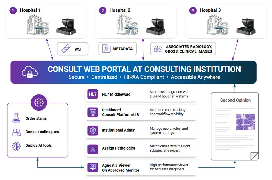
At PathPresenter, we recognized that remote consultation workflows require more than image viewing alone. They require infrastructure. That’s why we built a vendor-agnostic platform designed specifically for digital pathology consultations:

  • Integrated viewing of pathology, radiology, and clinical images.
  • Secure PHI-enabled collaboration
  • Structured consult workflows
  • Full audit trails
  • LIS integration
  • Async collaboration between institutions
  • Integrated viewing of pathology, radiology, and clinical images
Consult web portal diagram

PathPresenter has a secure, scalable, and HIPAA compliant digital workflow for clinical consults and referral

Most importantly, we designed the system to work across scanners, storage platforms, AI applications, and laboratory system because pathology is never a single-vendor environment.

Making Digitization Standard Operating Procedure

One of the biggest opportunities ahead is extending digital pathology beyond large academic centers and into outreach and referral hospitals.

A fully digital consultation workflow only works if cases can originate digitally. Historically, barriers like scanner costs, storage requirements, deployment complexity, and interoperability challenges slowed adoption. But cloud-native platforms, zero-footprint deployment models, and improved interoperability are rapidly changing the equation.

The transition to digital pathology will require collaboration across the industry. The good news is that this collaboration is finally beginning to happen, with many highly capable but lower cost scanners available.

The Future of Pathology Shouldn’t Depend on FedEx

Patients should not wait days or weeks for access to subspecialty expertise because slides are sitting in transit. The technology already exists to make pathology consultations digital, connected, and immediate.

Now it’s time for the industry to work together, and with partners at institutions and outreach hospitals to scale it.

Looking to Digitize your Consultation Workflow?

If your institution is thinking about expanding digital pathology consultations, enabling remote second opinions, or building a more connected pathology network, we’d love to talk. Reach out to the PathPresenter team to learn how we’re helping institutions modernize pathology workflows and expand access to subspecialty expertise worldwide.


About the Author

Patrick Myles CEO of PathPresenter. Previously, he was CEO of Huron Digital Pathology, and Vice President of Business Development for Teledyne DALSA. He served as a board member of the Digital Pathology Association.